Tools that allow neuroscientists to record and quantify functional activity within the living brain are in great demand. Traditionally, researchers have used techniques such as functional magnetic resonance imaging, but this method cannot record neural activity with high spatial resolution or in moving subjects. In recent years, a technology called optogenetics has shown considerable success in recording neural activity from animals in real time with single neuron resolution. Optogenetic tools use light to control neurons and record signals in tissues that are genetically modified to express light-sensitive and fluorescent proteins. However, existing technologies for imaging light signals from the brain have drawbacks in their size, imaging speed, or contrast that limit their applications in experimental neuroscience.
A technology called light-sheet fluorescence imaging shows promise for imaging brain activity in 3D with high speed and contrast (overcoming multiple limitations of other imaging technologies). In this technique, a thin sheet of laser light (light-sheet) is directed through a brain tissue region of interest, and fluorescent activity reporters within the brain tissues respond by emitting fluorescence signals that microscopes can detect. Scanning a light sheet in the tissue enables high-speed, high-contrast, volumetric imaging of the brain activity.
Currently, using light-sheet fluorescence brain imaging with nontransparent organisms (like a mouse) is difficult because of the size of the necessary apparatus. To make experiments with nontransparent animals and, in the future, freely moving animals feasible, researchers will first need to miniaturize many of the components.
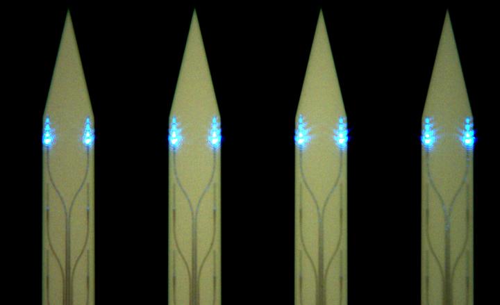
A key component for the miniaturization is the light-sheet generator itself, which needs to be inserted into the brain and thus must be as small as possible to avoid displacing too much brain tissue. In a new study reported in Neurophotonics, an international team of researchers from the California Institute of Technology (USA), University of Toronto (Canada), University Health Network (Canada), the Max Planck Institute of Microstructure Physics (Germany), and Advanced Micro Foundry (Singapore) developed a miniature light-sheet generator, or a photonic neural probe, that can be implanted into a living animal’s brain.
The researchers used nanophotonic technology to create ultrathin silicon-based photonic neural probes that emit multiple addressable thin sheets of light with thicknesses <16 micrometers over propagation distances of 300 micrometers in free space. When tested in brain tissues from mice that were genetically engineered to express fluorescent proteins in their brains, the probes permitted the researchers to image areas as large as 240 μm × 490 μm. Moreover, the level of image contrast was superior to that with an alternative imaging method called epifluorescence microscopy.
Describing the significance of his team’s work, the study’s lead author, Wesley Sacher, says, “This new implantable photonic neural probe technology for generating light sheets within the brain circumvents many of the constraints that have limited the use of light-sheet fluorescence imaging in experimental neuroscience. We predict that this technology will lead to new variants of light-sheet microscopy for deep brain imaging and behavior experiments with freely moving animals.”
Such variants would be a boon to neuroscientists seeking to understand the workings of the brain.
###
Read the open access research article by W.D. Sacher et al., “Implantable photonic neural probes for light-sheet fluorescence brain imaging,” Neurophotonics 8(2), 025003, doi 10.1117/1.NPh.8.2.025003.


