New imaging technique tracks traffic patterns of white blood cells

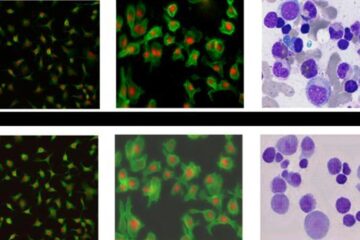
Microdomains are restricted areas on the surface of the cells in which receptors and signaling molecules accumulate during cell activation. Using digital multi-channel videomicroscopy, researchers were able to view white blood cell subsets and their forming microdomains in the vascular system in real time. In the upcoming March issue of Nature Methods, the new research displays the migration and inner workings of white blood cells in the small veins and bone marrow of mice.
“Intravital micropscopy (IVM) has contributed enormously to the recent mechanistic advances in leukocyte trafficking,” wrote Mount Sinai School of Medicine researchers lead author Elaine Y. Chiang, Andres Hidalgo, Jungshan Chang and Paul S. Frenette of the Department of Medicine’s Immunobiology Center and Black Family Stem Cell Institute in New York. “Multichannel digital fluorescence videomicropscopy vastly improves on prior intravital observations of leukocyte behavior in vivo by allowing the near-simultaneous observation of cell surface markers and microdomains.”
Researchers explored various mouse models including that of Sickle Cell disease which is a common inherited blood disorder in the United States, affecting 72,000 Americans according to the National Institutes of Health (NIH). Sickle Cell disease is a blood disorder that affects hemoglobin, a protein inside red blood cells that help carry oxygen throughout the body. Abnormal hemoglobin polymerizes, and red blood cells become sickle-shaped. Sickle-shaped red blood cells then clog blood vessels by sticking to white blood cells and depriving the body of needed oxygen.
Using this new imaging method, red blood cells in a mouse model of Sickle Cell disease were shown to interact specifically with neutrophils, a subset of inflammatory white blood cells. “The ease and rapidity of this method, and the abilty to combine it with current genetic labeling techniques will greatly aid investigations to elucidate in vivo imflammatory cell behavior, and provide a powerful tool for the development of therapeutics for these diseases,” according to Mount Sinai researchers.
“Sickle cell anemia was the first disease characterized at the molecular level but there is still no treatment for the acute vascular occlusions which are the hallmark of the disease,” said Mount Sinai researcher Dr. Paul S. Frenette. “In vivo imaging provides a window inside the body that allows us to see exactly which blood cells and which molecular constituents participate in the vascular occlusions. Advances in molecular imaging will likely lead to new targets for the treatment that will improve the life of patients afflicted by this terrible illness.”
Media Contact
More Information:
http://www.mssm.eduAll latest news from the category: Medical Engineering
The development of medical equipment, products and technical procedures is characterized by high research and development costs in a variety of fields related to the study of human medicine.
innovations-report provides informative and stimulating reports and articles on topics ranging from imaging processes, cell and tissue techniques, optical techniques, implants, orthopedic aids, clinical and medical office equipment, dialysis systems and x-ray/radiation monitoring devices to endoscopy, ultrasound, surgical techniques, and dental materials.
Newest articles

Bringing bio-inspired robots to life
Nebraska researcher Eric Markvicka gets NSF CAREER Award to pursue manufacture of novel materials for soft robotics and stretchable electronics. Engineers are increasingly eager to develop robots that mimic the…

Bella moths use poison to attract mates
Scientists are closer to finding out how. Pyrrolizidine alkaloids are as bitter and toxic as they are hard to pronounce. They’re produced by several different types of plants and are…

AI tool creates ‘synthetic’ images of cells
…for enhanced microscopy analysis. Observing individual cells through microscopes can reveal a range of important cell biological phenomena that frequently play a role in human diseases, but the process of…


