Motion pictures from living cells: Research team from Jena and Bielefeld improves superresolution microscopy
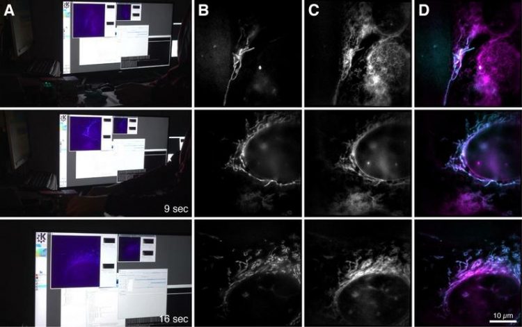
Images of the new microscope: The computer screen and the microscope images (right) show a bone cancer cell with mitochondria (blue) and endoplasmic reticulum (pink). Bielefeld University/ W. Hübner
This graphics card normally helps computer gamers to have a great gaming experience. The researchers, however, use it to observe the smallest cell components in action — in real time and with a very high frame rate.
“The image data can be reconstructed about twenty times faster than it would take on a PC,” explains Rainer Heintzmann of the Leibniz Institute of Photonic Technology (Leibniz IPHT), who laid the foundations for the process of structured illumination in high-resolution microscopy back in 1998. Together with him, the Bielefeld research team led by Prof. Thomas Huser further expanded the technology of Super-Resolved Structured Illumination Microscopy (SR-SIM).
In the fluorescence microscopic method SR-SIM, objects are irradiated with laser light using a special pattern. It excites special fluorescent molecules in the sample so that they emit light at a different wavelength. The microscopic image then shows this emitted light. It is first recorded in several individual images and then reconstructed as a high-resolution image on a computer.
“The second step in particular has taken a lot of time so far,” says Andreas Markwirth from the University of Bielefeld, the first author of the study. By using parallel computing methods on modern graphics cards for the new microscope, his team of researchers has now been able to significantly accelerate the image reconstruction process.
A minimum delay of 250 milliseconds is almost imperceptible to the human eye. The raw data can also be generated more quickly with the newly researched microscope.
Structures that are invisible to conventional microscopes
“This makes it possible to measure samples quickly and adapt test conditions immediately during an experiment instead of evaluating them afterwards,” says Rainer Heintzmann, describing the practical benefits of the new technology.
The scientists tested the method on biological cells and recorded the movements of mitochondria, the energy centres of the cells that are about one micrometer in size. “We were able to produce about 60 frames per second — a higher frame rate than those of motion pictures. The time between measurement and image is less than 250 milliseconds, which is why the technology allows real-time recordings,” says Andreas Markwirth.
So far, superresolution images have often been combined with conventional methods: A conventional fast microscope is used to first find structures. These structures can then be examined in detail using a superresolution microscope. “However, some structures are so small that they cannot be found with conventional microscopes, for example special pores in liver cells.
Our method is both high-resolution and fast, which enables biologists to investigate such structures,” said Thomas Huser. Another application for the new microscope is the investigation of viral particles on their way through the cell. “This enables us to understand exactly what happens during infection processes,” said Huser.
Superresolution microscopes have only been available for about 20 years. Ernst Abbe discovered in 1873 that the resolution of an optical system for visible light is limited to about 250 nanometres. In recent years, however, several optical methods have been developed in order to fall below Abbe's resolution limit. The Americans William E. Moerner and Eric Betzig, as well as the German Stefan Hell, were awarded the Nobel Prize in Chemistry in 2014 for developing a superresolution in the range of about 20 to 30 nanometers.
Prof. Dr. Rainer Heintzmann
Leibniz Institute of Photonic Technology (Leibniz IPHT)
and Institute for Physical Chemistry of the Friedrich-Schiller-University Jena
Head of the Microscopy Department at Leibniz-IPHT //Head of the Biomedical Imaging Working Group
+49 (0) 3641 · 206-431
rainer.heintzmann(a)leibniz-ipht.de
Andreas Markwirth, Mario Lachetta, Viola Mönkemöller, Rainer Heintzmann, Wolfgang Hübner, Thomas Huser, Marcel Müller: Video-rate multi-color structured illumination microscopy with simultaneous real-time reconstruction. Nature Communications, DOI 10.1038/s41467-019-12165-x, September 20, 2019.
Press release of the Bielefeld University on this topic: https://ekvv.uni-bielefeld.de/blog/pressemitteilungen/
Media Contact
All latest news from the category: Medical Engineering
The development of medical equipment, products and technical procedures is characterized by high research and development costs in a variety of fields related to the study of human medicine.
innovations-report provides informative and stimulating reports and articles on topics ranging from imaging processes, cell and tissue techniques, optical techniques, implants, orthopedic aids, clinical and medical office equipment, dialysis systems and x-ray/radiation monitoring devices to endoscopy, ultrasound, surgical techniques, and dental materials.


Newest articles

Properties of new materials for microchips
… can now be measured well. Reseachers of Delft University of Technology demonstrated measuring performance properties of ultrathin silicon membranes. Making ever smaller and more powerful chips requires new ultrathin…

Floating solar’s potential
… to support sustainable development by addressing climate, water, and energy goals holistically. A new study published this week in Nature Energy raises the potential for floating solar photovoltaics (FPV)…

Skyrmions move at record speeds
… a step towards the computing of the future. An international research team led by scientists from the CNRS1 has discovered that the magnetic nanobubbles2 known as skyrmions can be…

