Label-free intraoperative histopathology of surgical specimen
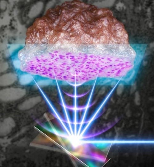
Using a MEMS scanner that can scan the light and sound waves quickly, high-speed pathological biopsy is performed on clinical tissue resected from cancer patients
Credit: POSTECH
Cancer diagnosis is confirmed through histopathology by removing a part of the tissue in question after conducting imaging tests such as MRI, CT, ultrasound, or endoscopy. Based on this clinical diagnosis, the cancerous tissue is surgically removed and suspected tissues or lymph nodes are additionally examined. Future treatment plans, chemotherapy, and radiation are formulated based on these results. Recently, a research team led by POSTECH and Gachon University College of Medicine has developed a machine learning-based histopathology method.
A research team led by Professor Chulhong Kim (POSTECH’s Department of Electrical Engineering, Department of IT Convergence Engineering, Department of Mechanical Engineering) in collaboration with researchers from Gachon University College of Medicine has developed a machine learning-based label-free histopathology device that can perform histopathology in real-time. The device avoids complicated procedures such as freezing, sectioning, or staining by using the ultraviolet (UV) photoacoustic imaging technology (UV-MEMS PAM) that converges ultra-high-speed MEMS scanner technology. This study was selected as the inside front cover paper in the September issue Laser and Photonics Reviews, an international scientific journal in the field of optics.
During cancer resection surgery, histopathologic examination is essential to confirm the site of the tumor. Frozen-section testing has been conventionally used to carry out the examination, but due to its complicated process, it can prolong surgery and potentially cause interpretation errors.
To overcome these shortcomings, the research team proposed a high-speed reflection-mode UV photoacoustic microscopy system using the 1-axis MEMS scanner as a novel label-free intraoperative histopathology method. The researchers succeeded in visualizing label-free cell nuclei of mouse and human tissues using this approach. In addition, by imaging clinical specimens resected from actual cancer patients and numerically quantifying the histopathologic results, the research team successfully demonstrated that the proposed UV-PAM system has great potential as an alternative intraoperative histopathology method.
Photoacoustic imaging not only enables three-dimensional imaging without an independent contrast agent, but also combines the advantages of both high-resolution optical imaging with the ultrasound imaging that can achieve structural and functional imaging of small cells and living tissues all the way to large organs. The microscope developed this time uses a high-speed MEMS scanner to significantly improve the imaging speed.
“The microscope developed in this study is new in that it distinguishes normal tissues from cancer tissues by performing photoacoustic histopathology on cancer tissues excised from actual cancer patients, and quantifying the pathological microstructures,” explained Professor Chulhong Kim of POSTECH. “This microscopy system is anticipated to dramatically cut down on the time it takes for biopsies during surgery, and to increase the stability and reliability of surgery and treatment.”
This research was conducted with the support from Korea’s Ministry of Science and ICT, the Ministry of Education, and the Ministry of Trade, Industry and Energy.
DOI: 10.1002/lpor.202100124
Article Title: Intraoperative Label-Free Photoacoustic Histopathology of Clinical Specimens
Article Publication Date: 16-Aug-2021
Media Contact
Jinyoung Huh
Pohang University of Science & Technology (POSTECH)
jyhuh@postech.ac.kr
Office: 82-54-279-2415
All latest news from the category: Medical Engineering
The development of medical equipment, products and technical procedures is characterized by high research and development costs in a variety of fields related to the study of human medicine.
innovations-report provides informative and stimulating reports and articles on topics ranging from imaging processes, cell and tissue techniques, optical techniques, implants, orthopedic aids, clinical and medical office equipment, dialysis systems and x-ray/radiation monitoring devices to endoscopy, ultrasound, surgical techniques, and dental materials.


Newest articles

High-energy-density aqueous battery based on halogen multi-electron transfer
Traditional non-aqueous lithium-ion batteries have a high energy density, but their safety is compromised due to the flammable organic electrolytes they utilize. Aqueous batteries use water as the solvent for…

First-ever combined heart pump and pig kidney transplant
…gives new hope to patient with terminal illness. Surgeons at NYU Langone Health performed the first-ever combined mechanical heart pump and gene-edited pig kidney transplant surgery in a 54-year-old woman…

Biophysics: Testing how well biomarkers work
LMU researchers have developed a method to determine how reliably target proteins can be labeled using super-resolution fluorescence microscopy. Modern microscopy techniques make it possible to examine the inner workings…

