Mapping the inner workings of a living cell
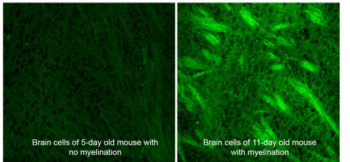
Using deuterium-labeled SRS imaging, researchers watched the brain cells of developing mice rapidly put on fat in a process called myelination. The ability to detect normal and abnormal myelination could help in detecting head injuries and monitoring the progression of multiple sclerosis. Credit: Min lab/Columbia
Imaging tools like X-rays and MRI have revolutionized medicine by giving doctors a close up view of the brain and other vital organs in living, breathing people. Now, Columbia University researchers report a new way to zoom in at the tiniest scales to track changes within individual cells.
Described in the latest issue of Nature Communications, the tool combines a widely used chemical tracer, D2O, or heavy water, with a relatively new laser-imaging method called stimulated Raman scattering (SRS). The technique's potential applications include helping surgeons quickly and precisely remove tumors, to helping to detect head injuries and developmental and metabolic disorders.
“We can use this technology to visualize metabolic activities in a wide range of animals,” said the study's senior author Wei Min, a chemistry professor at Columbia University. “By tracking where and when new proteins, lipids and DNA molecules are made, we can learn more about how animals develop and age, and what goes wrong in the case of injury and disease.”
The breakthrough involves the use of heavy water as a chemical tracer. Made by swapping water's hydrogen atoms with their heavier relative, deuterium, heavy water looks and tastes like regular water and in small doses (no more than five tablespoons for humans) is safe to drink. Once metabolized by cells in the body, heavy water is incorporated into newly made proteins, lipids and DNA, where the deuterium forms chemical bonds with carbon.
When these carbon-deuterium bonds are hit with light, they vibrate at varying frequencies, the researchers discovered, allowing each macromolecule to be identified as a protein, lipid or DNA. From these frequency signatures, they could track the growth of new proteins, lipids and DNA in the animal's brain, skin, gut and other organs.
Though heavy water is already used to label proteins and lipids to track metabolic changes, analysis is currently done on a mass spectrometer, on cells extracted from the body. This method now makes it possible to visualize subcellular changes in real time and space. “We get a continuous picture of what's happening inside living animal cells. Previously, we had only a snapshot,” said the study's co-lead author, Lingyan Shi, a postdoctoral researcher at Columbia.
In the study, the researchers diluted regular water with D2O and gave it to roundworms, mice and zebrafish embryos to drink. Aiming the SRS laser at a variety of tissue, they watched over hours and days as new deuterium-tagged proteins, lipids and DNA built up.
In one experiment, they watched a bright line emerge around fast-growing brain and colon tumors in the mice. As the cancerous cells divided, more deuterium was incorporated into their newly made proteins and lipids. “This method creates a sharp line between healthy and cancerous tissue, making it much easier to remove the tumor,” said Shi.
The experiments also offered new insights into cell development and aging.
*In the roundworm, they watched fat production rise and fall in the worm's reproductive system as it aged. Fat helps the worm's eggs to mature, and once this added fat was no longer useful, fat formation slowed, they found. They also saw clumps of new protein form in the older worm's body, suggesting that deuterium-labeled SRS imaging could be used to track protein deposits, and thus aging-related disease.
*In the developing brains of baby mice, they observed the formation of a layer of insulating fat, called the myelin sheath, around each cell. Watching the process in real time suggested to the researchers that deuterium-labeled SRS imaging could be used to tell if a child's brain is developing properly, or if patients suffering from multiple sclerosis, a disease that attacks the brain's myelin and disrupts information flow, might be recovering.
*In the sweat gland cells of mice, they watched as new lipids formed in cells at the outer edges of the sweat glands, pushing older cells inward. When those old cells finally reached the center of the glands, they died and were expelled in a process thought to moisturize the skin and hair above.
“The beauty of this mapping method is its simplicity,” says Eric Potma, a chemistry professor at University of California at Irvine who was not involved in the study. “It produces vivid images of metabolic activity in tissues with seemingly minimal effort. As the SRS microscope continues to get smaller, deuterium-labeled SRS imaging may help to catch tumors at much earlier stages.”
Acting on a hunch that the element hydrogen came in a heavier form, Harold Urey, then a chemistry professor at Columbia, succeeded in separating deuterium from liquid hydrogen in 1931. The discovery won him a Nobel Prize in chemistry three years later. In addition to serving as a tracer in mass spectroscopy, deuterium today is used to track changes in ocean circulation, study the formation of stars, and modulate chemical reactions in making nuclear power.
###
The other authors of the study are co-lead author Chaogu Zheng, Yihui Shen, Zhixing Chen, Edilson Silveira, Luyan Zhang, Mian Wei, Chang Liu, Carmen de Sena-Tomas and Kimara Targoff. The study was funded by the National Institute of General Medical Sciences, National Heart, Lung, and Blood Institute, National Institutes of Health, U.S. Army Research Office, Alfred P. Sloan Foundation and Camille and Henry Dreyfus Foundation.
Study: Optical imaging of metabolic dynamics in animals DOI 10.1038/s41467-018-05401-3
MEDIA CONTACT:
Kim Martineau klm32@columbia.edu 646-717-0134
SCIENTIST CONTACTS:
Wei Min
wm2256@columbia.edu
Lingyan Shi
ls3372@columbia.edu
Media Contact
More Information:
http://dx.doi.org/10.1038/s41467All latest news from the category: Life Sciences and Chemistry
Articles and reports from the Life Sciences and chemistry area deal with applied and basic research into modern biology, chemistry and human medicine.
Valuable information can be found on a range of life sciences fields including bacteriology, biochemistry, bionics, bioinformatics, biophysics, biotechnology, genetics, geobotany, human biology, marine biology, microbiology, molecular biology, cellular biology, zoology, bioinorganic chemistry, microchemistry and environmental chemistry.


Newest articles

Properties of new materials for microchips
… can now be measured well. Reseachers of Delft University of Technology demonstrated measuring performance properties of ultrathin silicon membranes. Making ever smaller and more powerful chips requires new ultrathin…

Floating solar’s potential
… to support sustainable development by addressing climate, water, and energy goals holistically. A new study published this week in Nature Energy raises the potential for floating solar photovoltaics (FPV)…

Skyrmions move at record speeds
… a step towards the computing of the future. An international research team led by scientists from the CNRS1 has discovered that the magnetic nanobubbles2 known as skyrmions can be…

