Lew lab sheds new light on cell membranes
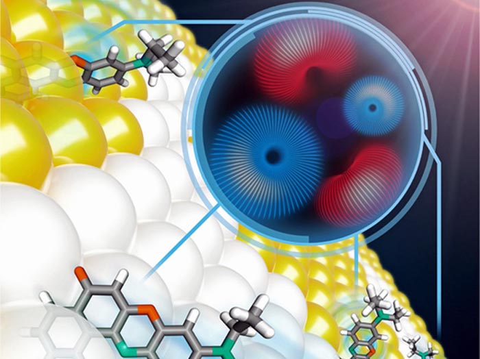
Polarized light in tandem with programmable microscopes allow researchers at the Lew Lab, at the McKelvey School of Engineering, to see both how molecules translate and how they rotate -- at the same time.
Credit: Oument Zhang/WashU
Researchers can now image cells, motions of molecules in 6D.
Research from the lab of Matthew Lew at Washington University in St. Louis offers entirely new ways to see the very small.
The research — two papers by PhD students at the McKelvey School of Engineering — was published in the journals Optica and Nano Letters.
They have developed novel hardware and algorithms that allow them to visualize the building blocks of the biological world beyond three dimensions in a way that, until now, wasn’t feasible. After all, cells are 3D objects and full of “stuff” — molecules — that moves around, rotates, spins and tumbles to drive life itself.
Like traditional microscopes, the work of two PhD students in the Lew lab, Tingting Wu and Oumeng Zhang, uses light to peer into the microscopic world — but their innovations are anything but traditional. Currently, when people use light in imaging, they are likely interested in how bright that light is or what color it is. But light has other properties, including polarization.
“Oumeng’s work twists the polarization of light,” said Lew, assistant professor in the Preston M. Green Department of Electrical & Systems Engineering. “This way, you can see both how things translate (move in straight lines) and rotate at the same time” — something traditional imaging doesn’t do.
“The development of new technology and the capability to see things we previously couldn’t see is exciting,” Zhang said. This unique capability to track both rotation and position at the same time gives him unique insights into how biological materials — human cells and pathogens, for instance — interact.
Wu’s research also provides a new way to image cell membranes and, in a way, to see inside of them. Using fluorescent tracer molecules, she maps how the tracers interact with fat and cholesterol molecules in the membrane, determining how the lipids are arranged and organized.
“Any cell membrane, any nucleus, anything in the cell is a 3D structure,” she said. “This helps us probe the full picture of a biological system. This enables us, for any biological sample, to see beyond three dimensions — we see the 3D structure plus three dimensions of molecular orientation, giving us 6D images.”
The researchers developed computational imaging technology, which synergizes software and hardware together, to successfully see the previously unseeable.
“That’s part of the innovation,” Lew said. “Traditionally, biological imaging labs have been tied down to whatever commercial manufacturers are making. But if we engineer things differently, we can do so much more.”
Journal: Optica
DOI: 10.1364/OPTICA.451899
Method of Research: Computational simulation/modeling
Subject of Research: Not applicable
Article Title: Dipole-spread-function engineering for simultaneously measuring the 3D orientations and 3D positions of fluorescent molecules
Article Publication Date: 20-May-2022
Media Contact
Brandie Jefferson
Washington University in St. Louis
brandie.jefferson@wustl.edu
Office: 314-935-5272
All latest news from the category: Life Sciences and Chemistry
Articles and reports from the Life Sciences and chemistry area deal with applied and basic research into modern biology, chemistry and human medicine.
Valuable information can be found on a range of life sciences fields including bacteriology, biochemistry, bionics, bioinformatics, biophysics, biotechnology, genetics, geobotany, human biology, marine biology, microbiology, molecular biology, cellular biology, zoology, bioinorganic chemistry, microchemistry and environmental chemistry.


Newest articles
Combatting disruptive ‘noise’ in quantum communication
In a significant milestone for quantum communication technology, an experiment has demonstrated how networks can be leveraged to combat disruptive ‘noise’ in quantum communications. The international effort led by researchers…
Stretchable quantum dot display
Intrinsically stretchable quantum dot-based light-emitting diodes achieved record-breaking performance. A team of South Korean scientists led by Professor KIM Dae-Hyeong of the Center for Nanoparticle Research within the Institute for…
Internet can achieve quantum speed with light saved as sound
Researchers at the University of Copenhagen’s Niels Bohr Institute have developed a new way to create quantum memory: A small drum can store data sent with light in its sonic…

