Cancer drugs: Interactive software simplifies research
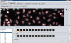
Zeta's clean graphical user interface: A few mouse clicks train the software to detect the cell cycle phases of HeLa cells. (c) Fraunhofer FIT<br>
Imaging technology is essential in the search for new active pharmaceutical ingredients for cancer drugs. Analyzing the large amounts of generated image data is time-consuming, often taking longer than the recording processes.
Here the Zeta software helps by simplifying the analysis and also by processing large amounts of data in a very short time. It takes just a few mouse clicks to train the software to recognize and classify specific cell patterns. Thus, Zeta can be adapted very flexibly to a wide range of biological analyses.
Fraunhofer FIT just extended their Zeta software to live cell imaging, which allows to monitor and record cancer cells across their full life cycle. Promising lead compounds are applied to the living cells – many thousands in parallel in different assays. Substances that block the division of cancerous cells but leave healthy cells untouched are potentials for cancer drugs.
These experiments are largely automated. A computer-controlled microscope generates series of images of the living cells, which are then processed by robust image analysis software. Here, the Zeta software assists the researchers by monitoring and quantifying the living cancer cells more precisely.
A particular challenge for the image analysis was to differentiate the phases of the cell cycle and to determine their transitions. “We need to identify the cells as objects and need to determine their individual cell cycle phases, but we also need to monitor the progression of the cycle from one phase to the next”, explains Prof. Thomas Berlage, the head of the Life Science Informatics department of Fraunhofer FIT.
A special feature of Zeta is its plug-in architecture, which allows the users to adapt the software very flexibly. When the program is started, a config file loads only those modules that are necessary for the analysis task at hand. As an example, the Foreground/Background plug-in is used to differentiate cell objects from the background. Here, Zeta offers an interactive procedure: A few mouse clicks by the user identify cancer cells and background regions. These examples train Zeta to discern cell objects and background; the software provides immediate visual feedback. This interactive trainability is a major advantage, as it enables Zeta to identify new cell types without changes to the software.
Additional plug-ins discern individual cells in cell clusters, identify their individual cell cycle phases or eliminate the motion artifacts typical of live cell imaging experiments.
The Fraunhofer Institute for Applied Information Technology FIT presents the Zeta software at Analytica 2012, held April 17 – 20, 2012 in Munich. Visit us in the Fraunhofer booth, Hall A1, Booth 433/530.
Contact:
Alex Deeg
pr@fit.fraunhofer.de
Telefon +49 2241 14-2208
Media Contact
More Information:
http://www.fit.fraunhofer.deAll latest news from the category: Trade Fair News


Newest articles

Superradiant atoms could push the boundaries of how precisely time can be measured
Superradiant atoms can help us measure time more precisely than ever. In a new study, researchers from the University of Copenhagen present a new method for measuring the time interval,…

Ion thermoelectric conversion devices for near room temperature
The electrode sheet of the thermoelectric device consists of ionic hydrogel, which is sandwiched between the electrodes to form, and the Prussian blue on the electrode undergoes a redox reaction…

Zap Energy achieves 37-million-degree temperatures in a compact device
New publication reports record electron temperatures for a small-scale, sheared-flow-stabilized Z-pinch fusion device. In the nine decades since humans first produced fusion reactions, only a few fusion technologies have demonstrated…

