Two-Colour Fluorescence Marker for Live Cell Imaging

Live Cell Imaging technologies provide the first detailed look at the dynamic structures and interactions of cellular organelles within living cells that may change within fractions of a second. A common problem to visualize different organelles is the necessity to use multiple fluorescent markers, which takes up a lot of time with additional costs. Scientists at the University of Göttingen have developed a powerful fluorescent marker for Live Cell Imaging to simultaneously study mitochondria as well as endosomes and lysosomes.
Further Information: PDF
MBM ScienceBridge GmbH
Phone: (0551) 30724-152
Contact
Dr. Jens-Peter Horst
Media Contact
All latest news from the category: Technology Offerings
Newest articles

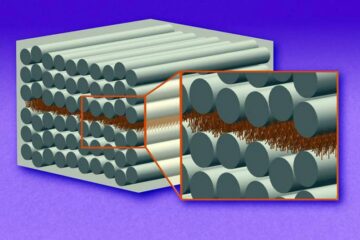
“Nanostitches” enable lighter and tougher composite materials
In research that may lead to next-generation airplanes and spacecraft, MIT engineers used carbon nanotubes to prevent cracking in multilayered composites. To save on fuel and reduce aircraft emissions, engineers…

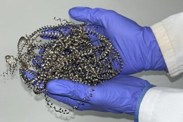
Trash to treasure
Researchers turn metal waste into catalyst for hydrogen. Scientists have found a way to transform metal waste into a highly efficient catalyst to make hydrogen from water, a discovery that…

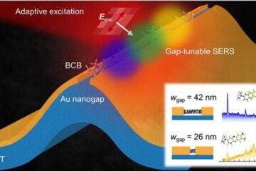
Real-time detection of infectious disease viruses
… by searching for molecular fingerprinting. A research team consisting of Professor Kyoung-Duck Park and Taeyoung Moon and Huitae Joo, PhD candidates, from the Department of Physics at Pohang University…


