New superlens opens door to nanoscale optical imaging, high-density optoelectronics
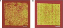
At left (A) is an image of an array of nanowires 60 nanometers wide created with the silver superlens. The center distance between each nanowire is 120 nanometers. To the right (B) is an image of the same nanowires. In this image, created without the superlens, the individual nanowires are not distinct. The scale bar on both images is 1 micrometer. (Image by Cheng Sun, UC Berkeley)
A group of scientists at the University of California, Berkeley, is giving new relevance to the term “sharper image” by creating a superlens that can overcome a limitation in physics that has historically constrained the resolution of optical images.
Using a thin film of silver as the lens and ultraviolet (UV) light, the researchers recorded the images of an array of nanowires and the word “NANO” onto an organic polymer at a resolution of about 60 nanometers. In comparison, current optical microscopes can only make out details down to one-tenth the diameter of a red blood cell, or about 400 nanometers.
The breakthrough, reported in the April 22 issue of the journal Science, opens the door to dramatic technological advances in nanoengineering that could eventually lead to DVDs that store the entire contents of the Library of Congress, and computer processors that can quickly search through such a huge volume of data, the researchers said.
“The field of optics is involved in much of today’s technology, including imaging and photolithography, which is used to make semiconductors and integrated circuits,” said Xiang Zhang, UC Berkeley associate professor of mechanical engineering and principal investigator of the study. “Our work has a far reaching impact on the development of detailed biomedical imaging, higher density electronic circuitry and ever-faster fiber optic communications.”
Nicholas Fang, one of Zhang’s former Ph.D. students and lead author of the paper, said a nearer term application would be the development of medical imaging devices that could reveal never-before-seen details with optical microscopy.
With current optical microscopes, scientists can only make out relatively large structures within a cell, such as its nucleus and mitochondria. With a superlens, optical microscopes could one day reveal the movements of individual proteins traveling along the microtubules that make up a cell’s skeleton, the researchers said.
Scanning electron and atomic force microscopes are now used to capture detail down to a few nanometers. However, such microscopes create images by scanning objects point by point, which means they are typically limited to non-living samples, and image capture times can take up to several minutes.
“Optical microscopes can capture an entire frame with a single snapshot in a fraction of a second,” said Fang, who is now an assistant professor of mechanical engineering at the University of Illinois at Urbana-Champaign. “That opens up nanoscale imaging to living materials, which can help biologists better understand cell structure and function in real time, and ultimately help in the development of new drugs to treat human diseases.”
The study is the latest entry in a hotly debated topic among physicists and engineers surrounding the creation of a lens that can break the so-called diffraction limit of optics through negative refraction.
Conventional lenses, whether manmade or natural, create images by capturing the propagating light waves all objects emit and then bending them. The angle of the bend is determined by the index of refraction and has always been positive.
Yet objects also emit “evanescent” waves that carry a great deal of detail but are far more elusive. Such evanescent waves decay exponentially and thus never make it to the image plane, an optics threshold known as the diffraction limit. Breaking this diffraction limit and capturing evanescent waves are critical to the creation of a 100-percent perfect representation of an object, considered the Holy Grail in optics.
In 2000, British physicist John Pendry theorized that a material capable of a negative index of refraction could capture and “refocus” evanescent waves into a perfect image. Pendry’s proposed “perfect lens” theory came more than 30 years after Russian physicist Victor Veselago first conceived of a negative refraction material that could reverse known optical phenomena.
These theories are based on the fact that when electromagnetic waves of light reach the surface of a negative refraction lens, they excite a collective movement of surface waves, such as electron oscillations, also known as surface plasmons. That results in an enhancement of the evanescent waves and is different from the way light typically behaves when it reaches a conventional lens.
Various negative refraction experiments were later conducted by researchers at UC San Diego, Boeing and Northeastern University, but they were limited to microwave beams.
In 2003, Zhang’s group was the first to confirm that optical evanescent waves are enhanced as they pass through a silver superlens in carefully designed conditions.
But it wasn’t until this latest experiment by Zhang’s group that optical imaging with a superlens was demonstrated. Zhang and his research team used UV light at a 365-nanometer wavelength in the new experiments, so the image created actually has more detail than is possible with beams in the microwave range.
The array of nanowires imaged measured 40 nanometers wide and the word NANO was about 60 nanometers wide. The objects, embedded onto a layer of chrome, were placed before the superlens, which was a layer of silver that was about 35 nanometers thick. The researchers recorded the image onto a photoresist, a polymer coating on the other side of the superlens that becomes insoluble when exposed to UV light.
“Our work provides a new imaging method that can beat the optical diffraction limit and that has tremendous potential to revolutionize a wide range of technologies,” said Zhang. “The key to the superlens is its ability to significantly enhance and recover the evanescent waves that carry information at very small scales. This enables imaging well below the diffraction limit.”
Notably, no lens is yet able to completely reconstitute all the evanescent waves emitted by an object, so the goal of a 100-percent perfect image is still out there. However, many scientists believe that a true perfect lens is not possible because there will always be some energy absorption loss as the waves pass through any known material.
“We did not create a perfect image in our experiment,” said Fang. “But it’s clear that our image is dramatically better than the one created without the silver superlens.”
In the long run, this line of research could lead to even higher resolution imaging for distant objects, the researchers said. This includes more detailed views of other planets as well as of human movement through surveillance satellites.
Media Contact
More Information:
http://www.berkeley.eduAll latest news from the category: Physics and Astronomy
This area deals with the fundamental laws and building blocks of nature and how they interact, the properties and the behavior of matter, and research into space and time and their structures.
innovations-report provides in-depth reports and articles on subjects such as astrophysics, laser technologies, nuclear, quantum, particle and solid-state physics, nanotechnologies, planetary research and findings (Mars, Venus) and developments related to the Hubble Telescope.


Newest articles

“Nanostitches” enable lighter and tougher composite materials
In research that may lead to next-generation airplanes and spacecraft, MIT engineers used carbon nanotubes to prevent cracking in multilayered composites. To save on fuel and reduce aircraft emissions, engineers…

Trash to treasure
Researchers turn metal waste into catalyst for hydrogen. Scientists have found a way to transform metal waste into a highly efficient catalyst to make hydrogen from water, a discovery that…

Real-time detection of infectious disease viruses
… by searching for molecular fingerprinting. A research team consisting of Professor Kyoung-Duck Park and Taeyoung Moon and Huitae Joo, PhD candidates, from the Department of Physics at Pohang University…

