Microscopes provide new view for tissue engineering
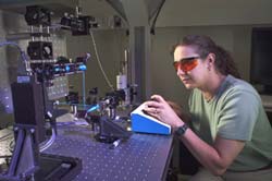
NIST physical scientist Joy Dunkers positions a polymer scaffold sample for imaging. <br>©Robert Rathe
In the November issue of Optics Express*, National Institute of Standards and Technology (NIST) scientists describe a novel combination of microscopes that can peer deep into tissue-engineering scaffolds and monitor the growth and differentiation of cells ultimately intended to develop into implantable organs or other body-part replacements.
The new dual-imaging tool provides a much needed capability for the emerging tissue engineering field, which aims to regenerate form and function in damaged or diseased tissues and organs. Until now, scrutiny of this complicated, three-dimensional process has been limited to the top-most layers of the scaffolds used to coax and sustain cell development.
Composed of biodegradable polymers or other building materials, scaffolds are seeded with cells that grow, multiply, and assemble into three-dimensional tissues. Whether the cells respond and organize as intended in this synthetic environment depends greatly on the composition, properties, and architecture of the scaffolds’ porous interiors. Tools for simultaneously monitoring microstructure and cellular activity can help scientists to tease apart the essentials of this interactive relationship. In turn, such knowledge can speed development of tissue-engineered products ranging from skin replacements to substitute livers to inside-the-body treatments of osteoporosis.
NIST scientist Joy Dunkers and her colleagues paired an optical coherence microscope – a high-resolution probe of the scaffold interior – with a confocal fluorescence microscope – used to track cells stained with a fluorescent dye. The instruments provide simultaneous images that can be merged to create a comprehensive rendering of microstructure and cellular activity. By stacking the sectional images, they can create a top-to-bottom movie showing structural and cellular details throughout the scaffold’s volume.
*J. P. Dunkers, M. T. Cicerone, and N. R. Washburn, “Collinear optical coherence and confocal fluorescence microscopies for tissue engineering,” Optics Express, Vol. 11, No. 23, pp. 3074-3079. [http://www.opticsexpress.org].
Media Contact
All latest news from the category: Physics and Astronomy
This area deals with the fundamental laws and building blocks of nature and how they interact, the properties and the behavior of matter, and research into space and time and their structures.
innovations-report provides in-depth reports and articles on subjects such as astrophysics, laser technologies, nuclear, quantum, particle and solid-state physics, nanotechnologies, planetary research and findings (Mars, Venus) and developments related to the Hubble Telescope.


Newest articles

Superradiant atoms could push the boundaries of how precisely time can be measured
Superradiant atoms can help us measure time more precisely than ever. In a new study, researchers from the University of Copenhagen present a new method for measuring the time interval,…

Ion thermoelectric conversion devices for near room temperature
The electrode sheet of the thermoelectric device consists of ionic hydrogel, which is sandwiched between the electrodes to form, and the Prussian blue on the electrode undergoes a redox reaction…

Zap Energy achieves 37-million-degree temperatures in a compact device
New publication reports record electron temperatures for a small-scale, sheared-flow-stabilized Z-pinch fusion device. In the nine decades since humans first produced fusion reactions, only a few fusion technologies have demonstrated…

