Mini X-ray source with laser light
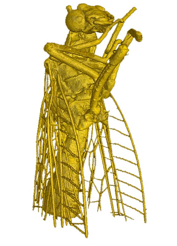
The world’s first image of a fly taken with the help of a all-laser-based X-ray tomography imaging method. It consists of around 1500 individual images. Photo: Karsch/Pfeiffer
With laser light, physicists in Munich have built a miniature X-ray source. In so doing, the researchers from the Laboratory of Attosecond Physics of the Max Planck Institute of Quantum Optics and the Technische Universität München (TUM) captured three-dimensional images of ultrafine structures in the body of a living organism for the first time with the help of laser-generated X-rays.
Using light-generated radiation combined with phase-contrast X-ray tomography, the scientists visualized ultrafine details of a fly measuring just a few millimeters. Until now, such radiation could only be produced in expensive ring accelerators measuring several kilometers in diameter. By contrast, the laser-driven system in combination with phase-contrast X-ray tomography only requires a university laboratory to view soft tissues. The new imaging method could make future medical applications more cost-effective and space-efficient than is possible with today’s technologies.
When the physicists Prof. Stefan Karsch and Prof. Franz Pfeiffer illuminate a tiny fly with X-rays, the resulting image captures even the finest hairs on the wings of the insect. The experiment is a pioneering achievement. For the first time, scientists coupled their technique for generating X-rays from laser pulses with phase-contrast X-ray tomography to visualize tissues in organisms. The result is a three-dimensional view of the insect in unprecedented detail.
The X-rays required were generated by electrons that were accelerated to nearly the speed of light over a distance of approximately one centimeter by laser pulses lasting around 25 femtoseconds. A femtosecond is one millionth of a billionth of a second. The laser pulses have a power of approximately 80 terawatts (80 x 10^12 watts). By way of comparison: an atomic power plant generates 1,500 megawatts (1.5 x 10^9 Watt).
First, the laser pulse ploughs through a plasma consisting of positively charged atomic cores and their electrons like a ship through water, producing a wake of oscillating electrons. This electron wave creates a trailing wave-shaped electric field structure on which the electrons surf and are accelerated in the process. The particles then start to vibrate, emitting X-rays. Each light pulse generates an X-ray pulse. The X-rays generated have special properties: They have a wavelength of approximately 0.1 nanometers, a duration of only about five femtoseconds, and are spatially coherent, i.e. they appear to come from a point source.
For the first time, the researchers combined their laser-driven X-rays with a phase-contrast imaging method developed by a team headed by Prof. Franz Pfeiffer of the TUM. Instead of the usual absorption of radiation, they used X-ray refraction to accurately image the shapes of objects, including soft tissues. For this to work, the spatial coherence mentioned above is essential.
This laser-based imaging technique enables the researchers to view structures around one tenth to one hundredth the diameter of a human hair. Another advantage is the ability to create three-dimensional images of objects. After each X-ray pulse, meaning after each frame, the specimen is rotated slightly. For example, about 1,500 individual images were taken of the fly, which were then assembled to form a 3D data set.
Due to the shortness of the X-ray pulses, this technique may be used in future to freeze ultrafast processes on the femtosecond time scale e.g. in molecules – as if they were illuminated by a femtosecond flashbulb.
The technology is particularly interesting for medical applications, as it is able to distinguish between differences in tissue density. Cancer tissue, for example, is less dense than healthy tissue. The method therefore opens up the prospect of detecting tumors that are less than one millimeter in diameter in an early stage of growth before they spread through the body and exert their lethal effect. For this purpose, however, researchers must shorten the wavelength of the X-rays even further in order to penetrate thicker tissue layers.
Thorsten Naeser
Original publication:
J. Wenz, S. Schleede, K. Khrennikov, M. Bech, P. Thibault, M. Heigoldt, F. Pfeiffer und S. Karsch, Quantitative X-ray phase-contrast microtomography from a compact laser-driven betatron source
Nature Communications, 20 July 2015, doi: 10.1038/ncomms8568
Further information can be obtained from:
Prof. Dr. Stefan Karsch
Faculty of Physics at Ludwig-Maximilians-Universität Munich
Am Coulombwall 1, 85748 Garching
Tel.: 089 32905 242
E-mail: stefan.karsch@mpq.mpg.de
www.attoworld.de , www.lex-photonics.de
Prof. Franz Pfeiffer
TUM, Chair of Biomedical Physics
James-Franck-Str. 1, 85748 Garching bei München
Tel.: 089 289 10807
E-mail: franz.pfeiffer@tum.de
Media Contact
All latest news from the category: Physics and Astronomy
This area deals with the fundamental laws and building blocks of nature and how they interact, the properties and the behavior of matter, and research into space and time and their structures.
innovations-report provides in-depth reports and articles on subjects such as astrophysics, laser technologies, nuclear, quantum, particle and solid-state physics, nanotechnologies, planetary research and findings (Mars, Venus) and developments related to the Hubble Telescope.


Newest articles

Silicon Carbide Innovation Alliance to drive industrial-scale semiconductor work
Known for its ability to withstand extreme environments and high voltages, silicon carbide (SiC) is a semiconducting material made up of silicon and carbon atoms arranged into crystals that is…

New SPECT/CT technique shows impressive biomarker identification
…offers increased access for prostate cancer patients. A novel SPECT/CT acquisition method can accurately detect radiopharmaceutical biodistribution in a convenient manner for prostate cancer patients, opening the door for more…

How 3D printers can give robots a soft touch
Soft skin coverings and touch sensors have emerged as a promising feature for robots that are both safer and more intuitive for human interaction, but they are expensive and difficult…

