Virginia Tech engineers introduce thermotherapy as a chemotherapy alternative

The cancer treatment uses hyperthermia to elevate the temperature of tumor cells, while keeping the surrounding healthy tissue at a lower degree of body heat. The investigators used both in vitro and in vivo experiments to confirm their findings.
The collaborators are Monrudee Liangruksa, a Virginia Tech graduate student in engineering science and mechanics, and her thesis adviser, Ishwar Puri, professor and head of the department, along with Ranjan Ganguly of the department of power engineering at Iadavpur Univesity, Kolkata, India.
Liangruska of Bangkok, Thailand, presented the paper at the meeting.
In an interview prior to the presentation, Puri explained that to further perfect the technique they used ferrofluids to induce the hyperthermia. A ferrofluid is a liquid that becomes strongly magnetized in the presence of a magnetic field. The magnetic nanoparticles are suspended in the non-polar state.
“These fluids can then be magnetically targeted to cancerous tissues after intravenous application,” Puri said. “The magnetic nanoparticles, each billionths of a meter in size, seep into the tissue of the tumor cell due to the high permeability of these vessels.”
Afterwards, the magnetic nanoparticles are heated by exposing the tumor to a high frequency alternating magnetic field, causing the tissue's death by heating. This process is called magnetic fluid hyperthermia and they have nicknamed it thermotherapy.
Temperatures in the range of 41 to 45 degrees Celsius are enough to slow or halt the growth of cancerous tissue. However, without the process of magnetic fluid hyperthermia, these temperatures also destroy healthy cells.
“The ideal hyperthermia treatment sufficiently increases the temperature of the tumor cells for about 30 minutes while maintaining the healthy tissue temperature below 41 degrees Celsius,” Puri said. “Our ferrofluid-based thermotherapy can be also accomplished through thermoablation, which typically heats tissues up to 56 degrees C to cause their death, coagulation, or carbonization by exposure to a noninvasive radio frequency, alternating current magnetic field. Local heat transfer from the nanoparticles increases the tissue temperature and ruptures the cell membranes.”
Puri added that testing showed iron oxide nanoparticles are “the most biocompatible agents for magnetic fluid hyperthermia.” Platinum and nickel also act as magnetic nanoparticles but they “are toxic and vulnerable” when exposed to oxygen.
The researchers plan to test their analytical approach by conducting experiments on various cancer cells in collaboration with Dr. Elankumaran Subbiah of the Virginia-Maryland School of Veterinary Medicine. A senior design team consisting of five engineering science and mechanics undergraduate Virginia Tech students is fabricating an apparatus for these tests.
Media Contact
More Information:
http://www.vt.eduAll latest news from the category: Health and Medicine
This subject area encompasses research and studies in the field of human medicine.
Among the wide-ranging list of topics covered here are anesthesiology, anatomy, surgery, human genetics, hygiene and environmental medicine, internal medicine, neurology, pharmacology, physiology, urology and dental medicine.
Newest articles

Silicon Carbide Innovation Alliance to drive industrial-scale semiconductor work
Known for its ability to withstand extreme environments and high voltages, silicon carbide (SiC) is a semiconducting material made up of silicon and carbon atoms arranged into crystals that is…

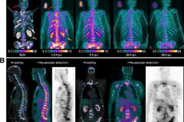
New SPECT/CT technique shows impressive biomarker identification
…offers increased access for prostate cancer patients. A novel SPECT/CT acquisition method can accurately detect radiopharmaceutical biodistribution in a convenient manner for prostate cancer patients, opening the door for more…

How 3D printers can give robots a soft touch
Soft skin coverings and touch sensors have emerged as a promising feature for robots that are both safer and more intuitive for human interaction, but they are expensive and difficult…


