Testing corneal cell quality? Apply physics
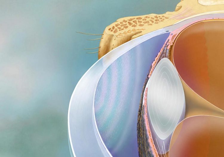
This is a cutaway view of a human eye, showing the honeycomb-like arrangement of corneal endothelial cells. Credit: Kyoto University/Tomo Narashima
Our eyes — the windows to the soul — need constant care, and as we age, they sometimes also need significant repair.
The panes of these windows — the corneas — are transparent tissues that have been the focus of some of the oldest and most common transplantation surgeries. Now thanks to researchers in Kyoto, some such transplants may become even safer.
The team, led by Kyoto University physicists and Kyoto Prefectural University of Medicine (KPUM) ophthalmologists, has developed a 'quantitative biomarker' that makes it possible to assess the quality of corneal cells — and even predict their long-term efficacy — through simple observation. A report on their findings appeared recently in Nature Biomedical Engineering.
“Cornea transplantations become necessary when 'corneal endothelial cells' decrease in number, resulting in haziness,” explains project leader Motomu Tanaka.
Endothelia don't multiply well in the human body, which is why there has been a need to rely on the transplantation of donor corneas for treatment. Fortunately, in 2009 a team of ophthalmologists at KPUM succeeded in developing a method to culture the cells in a dish.
“These new cells could then be then transplanted into the eyes of patients and restore their corneas to health,” says KPUM's Morio Ueno.
This method has shown significant promise in clinical trials, but two major obstacles to wider application remain: quality control of cells before injection and confirmation of long-term functionality.
Typically, cell quality is assessed through protein expression patterns via 'flow cytometry'. However, a single test requires almost 100,000 cells and relies heavily on the observations and experience of senior professionals.
“Cells in a tissue are constantly interacting with each other to maintain a steady state, called homeostasis,” explains first author Akihisa Yamamoto, adding that the concept of 'colloid physics' — a method for measuring interactions of micro- and nanoparticles — was employed to assess the cornea cells.
“Calculating the interactions between all cells in the cornea allowed us to find the 'spring constant', correlating with collective cell order.”
Assessment is relatively simple. Researchers only need to extract the 'rims' of the cells, either from a microscopic image of the cells in a culture dish or from ophthalmological inspection images of the patients' eyes. Both the quality of the cells and their long-term efficacy can be determined with just one equation.
The procedure has potential applications in preemptive medicine, enabling clinicians and doctors to intervene before more severe symptoms appear.
“Our results are thanks to the united effort of physicists and doctors engaged in regenerative medicine,” concludes Tanaka. “We foresee that our 'quantitative biomarker', and the concept behind it, will be applied to other epithelial cell cultures and tissues in the future.”
###
The paper “A physical biomarker of the quality of cultured corneal endothelial cells and of the long-term prognosis of corneal restoration in patients” appeared on 22 July 2019 in Nature Biomedical Engineering, with doi: 10.1038/s41551-019-0429-9
About Kyoto University
Kyoto University is one of Japan and Asia's premier research institutions, founded in 1897 and responsible for producing numerous Nobel laureates and winners of other prestigious international prizes. A broad curriculum across the arts and sciences at both undergraduate and graduate levels is complemented by numerous research centers, as well as facilities and offices around Japan and the world. For more information please see: http://www.
Media Contact
More Information:
http://dx.doi.org/10.1038/s41551-019-0429-9All latest news from the category: Health and Medicine
This subject area encompasses research and studies in the field of human medicine.
Among the wide-ranging list of topics covered here are anesthesiology, anatomy, surgery, human genetics, hygiene and environmental medicine, internal medicine, neurology, pharmacology, physiology, urology and dental medicine.


Newest articles

Bringing bio-inspired robots to life
Nebraska researcher Eric Markvicka gets NSF CAREER Award to pursue manufacture of novel materials for soft robotics and stretchable electronics. Engineers are increasingly eager to develop robots that mimic the…

Bella moths use poison to attract mates
Scientists are closer to finding out how. Pyrrolizidine alkaloids are as bitter and toxic as they are hard to pronounce. They’re produced by several different types of plants and are…

AI tool creates ‘synthetic’ images of cells
…for enhanced microscopy analysis. Observing individual cells through microscopes can reveal a range of important cell biological phenomena that frequently play a role in human diseases, but the process of…

