Artery variations increase complication risk in liver transplants

3D MDCT accurate for imaging liver arteries
3D MDCT angiography is a more efficient way to classify liver arterial anatomy before liver surgery, according to researchers from Duke University in Durham, NC. In the study, 43 patients were evaluated before using 3D MDCT angiography. In 40 of 43 patients, surgical findings concurred with MDCT findings, indicating that 3D MDCT is an accurate method for imaging liver arteries prior to surgery, said Erik K. Paulson, MD, an author on the paper.
“Previously, patients underwent catheter angiography, diagnostic CT and sometimes an MRI as well. Now, all the relevant imaging issues can be addressed in one noninvasive test,” said Dr. Paulson “This saves cost, reduces risk to the patient and saves time,” he said.
In a separate study, University of Iowa researchers have found that using MR angiography before a liver transplant can help identify which patients will suffer complications.
The study found that if the liver artery anatomy is unusual, there is an increased risk of complications after the transplant procedure, said Kousei Ishigami, MD, lead author of the paper. The study analyzed 84 patients who underwent the imaging prior to liver transplantation. Of the sixty patients who had normal artery anatomy, only two had complications after surgery, whereas five of the 24 with variant anatomy experienced complications after surgery.
According to the authors of the study, having such a variant anatomy, which occurs during fetal development, usually does not affect a healthy person. However, the knowledge can be of great help for some liver transplant recipients. “If we know a transplant recipient has a variant liver artery, we need to carefully follow these patients after liver transplantation. Some transplant surgeons may also want to know the recipient’s liver arterial anatomy before transplantation since knowledge of it may change the surgeon’s technique or plan,” said Dr. Ishigami, now at Kyushu University in Japan. “It should be noted, though, that MR angiography is impractical in emergency situations or when a patient is unable to remain still or hold their breath for the exam,” he added.
Both studies appear in the December 2004 issue of the American Journal of Roentgenology.
Media Contact
More Information:
http://www.arrs.orgAll latest news from the category: Health and Medicine
This subject area encompasses research and studies in the field of human medicine.
Among the wide-ranging list of topics covered here are anesthesiology, anatomy, surgery, human genetics, hygiene and environmental medicine, internal medicine, neurology, pharmacology, physiology, urology and dental medicine.
Newest articles

Properties of new materials for microchips
… can now be measured well. Reseachers of Delft University of Technology demonstrated measuring performance properties of ultrathin silicon membranes. Making ever smaller and more powerful chips requires new ultrathin…

Floating solar’s potential
… to support sustainable development by addressing climate, water, and energy goals holistically. A new study published this week in Nature Energy raises the potential for floating solar photovoltaics (FPV)…

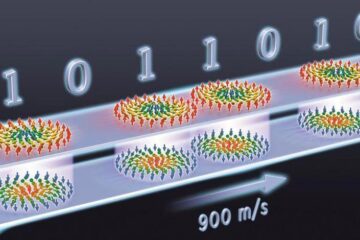
Skyrmions move at record speeds
… a step towards the computing of the future. An international research team led by scientists from the CNRS1 has discovered that the magnetic nanobubbles2 known as skyrmions can be…


