License for self-destruction: it is in the lung that Tcells obtain the capacity to attack the brain
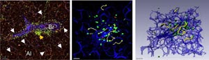
The lung as niche for T cells. The pictures show autoaggressive T cells (green, arrows) accumulating in the lung. Here they gain the ability to migrate to their target tissue, the central nervous system. The cells accumulate in specialized lymphatic structures (BALT, yellow arrow) and move along the outer surfaces and within the airways (bronchial tubes, Br), which they use as a type of road. Some cells also move through the air sacs (alveoli, Al). Fig. left: Lung explant as viewed with 2-photon microscopy. Fig. center: 10-minute time projection. Red dots: stationary T cells, yellow lines show the path taken by motile T cells. Fig. right: 3-D reconstruction. Source: umg/imsf göttingen<br>
Autoimmune diseases are triggered by immune cells that attack the body’s own tissue. In multiple sclerosis (MS) immune cells succeed in invading nervous tissue and sparking off a destructive inflammation there which can be accompanied by neurological deficits such as paralysis and somatosensory defects.
A healthy brain is practically free from immune cells, because the nervous system is separated from the rest of the body via specialized blood vessels that prevent immune cells from entering it from the blood.
Up to now it has been unclear how in MS immune cells can overcome this barrier and seemingly pass unhindered into the brain tissue. A research team under the direction of Prof. A. Flügel, head of the Department of Neuroimmunology and the Institute for Multiple Sclerosis Research (IMSF), University Medical Center Göttingen, could now show that these disease-causing immune cells are programmed in the lung to be more motile and to efficiently break through blood vessel barriers. The team’s results were published end of August 2012 in the international journal “NATURE”. The research was supported in part by an individual project grant from the Hertie Foundation, which also funds the IMSF.
Original publication: Francesca Odoardi, Christopher Sie, Kristina Streyl, Vijay K. Ulaganathan, Christian Schläger, Dmitri Lodygin, Klaus Heckelsmiller, Wilfried Nietfeld, Joachim Ellwart, Wolfgang E.F. Klinkert, Claudio Lottaz, Mikhail Nosov, Volker Brinkmann, Rainer Spang, Hans Lehrach, Martin Vingron, Hartmut Wekerle, Cassandra Flügel-Koch and Alexander Flügel. T cells become licensed in the lung to enter the central nervous system, Nature (2012) 488: 675-679.
Specialized immune cells, so-called T cells, are held to be the cause of MS. Even though nearly every healthy human harbors potentially disease-causing T cells in his or her immune system, only around 0.1% of the population actually develops a manif-est MS. One of the reasons for this is that normally T cells are stopped from entering the brain by a virtually impermeable vascular barrier separating the central nervous system from the blood circulation. “Earlier work in experimental MS research showed that when T cells are pre-activated outside nervous tissue they are very well able to pass into the brain and trigger MS-like symptoms there”, explains Alexander Flügel. “However, we wanted to find out exactly where in the body these T cells are activated and exactly which properties enable them to overcome the blood–brain barrier.”
ACCESS CODE TO THE BRAIN
Scientists working at the University Medical Center Göttingen initially discovered that disease-causing T cells cannot enter the brain immediately after activation but rather have to “learn” how to do so. During this learning process the T cells completely re-gear themselves. They stop dividing and throttle their production of proteins that fo-ment inflammation. Instead they are programmed for migration: they become more motile, and specialized receptors appear on their surface membranes. These recep-tors are like little antennae that enable a T cell to orientate itself by picking up signals from its environment and to cleave to surfaces.
The Göttingen scientists discovered that the receptor Ninjurin 1, previously unknown to have any relevance to T cells, controls the ability of T cells to cleave to the inner side of the brain’s blood vessels and thus is of great significance for the migration of T cells from the blood into the nervous tissue. Once the T cells arrive in the nervous tissue, this program goes into reverse: the immigrant T cells are reactivated and they produce inflammatory mediators that set the tissue-damaging autoimmune processes in motion typical to MS.
THE LUNG AS SWITCHING STATION AND DEPOT
But where in the body are the T cells programmed for migration? The Göttingen scien-tists could also make new, unexpected discoveries on this question. They found out that activated T cells migrate directly from the circulation into the lung. Once in the lung tissue the cells move forward with increasing speed along its blood vessels and airways to reach the adjoining lymph nodes from where they re-enter the blood circu-lation and spleen and ultimately invade the nervous system. Curiously, when in the lung the T cells do not only creep along the outer surface of the bronchial tubes but also crawl briskly along the inner surfaces of the airways where breathing air is circu-lated. Using a special microscopic technique the researchers could observe in living lung tissue the T cells using the bronchial tubes as a kind of highway. And indeed, when activated T cells are introduced directly into the airways they are able to set an autoimmune disease process in motion. It is also here in the lung where the first deci-sive steps take place towards programming the disease-causing T cells into a migratory mode.
OUTLOOK: THE LUNG AS A POSSIBLE POINT OF ORIGIN FOR MS ATTACKS
The direct relevance of these results to the human disease MS lies in the possibility that infections of the respiratory tract and/or lung irritants, e.g. smoking, can trigger disease attacks. The scientists of this study discovered potentially autoaggressive T cells dwelling long-term in the lung as immunological memory cells. When stimulated locally, these “sleeping” cells became active: They migrated to the brain and triggered off an MS-type disease there.
The key role of the lung in activating and reprogramming disease-causing T cells could also be valid for other organ systems such as the gut or urinary tract, though perhaps less dramatically. Recent comprehensive genetic analyses could identify various genes in persons suffering from MS that made these persons more suscepti-ble to the disease. “Interestingly, a significant number of these genes were the same as those found in the current study to be involved in the migratory programming of T cells”, says Alexander Flügel. The aim of further studies will therefore be to find genes from the migratory programming that are suitable as therapeutic targets.
FURTHER INFORMATION:
University Medical Center Göttingen, Georg-August University
Department of Neuroimmunology / Institute for Multiple Sclerosis Research
Alexander Flügel, MD, Phone +49 (0) 551 / 39-13332, IMSF@med.uni-goettingen.de
Media Contact
More Information:
http://www.uni-goettingen.deAll latest news from the category: Health and Medicine
This subject area encompasses research and studies in the field of human medicine.
Among the wide-ranging list of topics covered here are anesthesiology, anatomy, surgery, human genetics, hygiene and environmental medicine, internal medicine, neurology, pharmacology, physiology, urology and dental medicine.


Newest articles

High-energy-density aqueous battery based on halogen multi-electron transfer
Traditional non-aqueous lithium-ion batteries have a high energy density, but their safety is compromised due to the flammable organic electrolytes they utilize. Aqueous batteries use water as the solvent for…

First-ever combined heart pump and pig kidney transplant
…gives new hope to patient with terminal illness. Surgeons at NYU Langone Health performed the first-ever combined mechanical heart pump and gene-edited pig kidney transplant surgery in a 54-year-old woman…

Biophysics: Testing how well biomarkers work
LMU researchers have developed a method to determine how reliably target proteins can be labeled using super-resolution fluorescence microscopy. Modern microscopy techniques make it possible to examine the inner workings…

