Uncovering cancer's inner workings by capturing live images of growing tumors
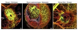
The new imaging tool reveals strikingly different networks of blood vessels surrounding different types of tumors in a mouse model. Left: breast cancer in the breast. Middle: metastatic breast cancer in the brain. Right: ectopic breast cancer in the skin.<br><br>Credit: Nature Medicine<br>
Scientists seeking new ways to fight cancer often try to understand the subtle, often invisible, changes to DNA, proteins, cells, and tissue that alter the body's normal biology and cause disease.
Now, to aid in that fight, a team of researchers has developed a sophisticated new optical imaging tool that enables scientists to look deep within tumors and uncover their inner workings. In experiments that will be described at Frontiers in Optics (FiO), The Optical Society's (OSA) Annual Meeting, Dai Fukumura and his colleagues will present new optical imaging techniques to track the movement of molecules, cells, and fluids within tumors; examine abnormalities in the blood vessel network inside them; and observe how the tumors were affected by treatments.
These techniques, created by Fukumura and his long-term collaborators at Massachusetts General Hospital and Harvard Medical School, combine two different high-tech optical imaging methods that were custom-built for the research. One is called multiphoton laser-scanning microscopy (MPLSM), which is an advanced fluorescence imaging technology that is now commercially available at the high end of the microscope market. The other is called optical frequency domain imaging (OFDI), which images tissues by their light scattering properties. According to Fukumura, OFDI is gaining popularity in the optical imaging field but has yet to become commercially available.
“MPLSM overcomes many of the limitations from which conventional microscopy and confocal microscopy suffer, and OFDI provides robust large volume imaging data,” Fukumura said.
Fukumura will present their research at FiO 2013, taking place Oct. 6-10 in Orlando, Fla. There, he will describe how his unique technique can image tumors inside and out, and show detailed pictures of live tumors—images that he and colleagues call “astonishing.”
He added that while the new combined approach would be too expensive to be used for routine diagnostic purposes, it promises to help researchers better understand the intricate workings of human cancer and aid in drug discovery to treat cancer. “These optical imaging approaches can provide unprecedented insights in the biology and mechanisms of cancer,” he said.
Presentation FW5A.2, “Experimental Methods for In Vivo Tissue Imaging,” takes place Wednesday, Oct. 9 at 4:15 p.m. EDT at the Bonnet Creek Ballroom, Salon IV at the Hilton Bonnet Creek in Orlando, Fla.
About the Meeting
Frontiers in Optics (FiO) 2013 is The Optical Society's (OSA) 97th Annual Meeting and is being held together with Laser Science XXIX, the annual meeting of the American Physical Society (APS) Division of Laser Science (DLS). The two meetings unite the OSA and APS communities for five days of quality, cutting-edge presentations, fascinating invited speakers and a variety of special events spanning a broad range of topics in optics and photonics—the science of light—across the disciplines of physics, biology and chemistry. An exhibit floor featuring leading optics companies will further enhance the meeting. More information at http://www.FrontiersinOptics.org.
About OSA
Founded in 1916, The Optical Society (OSA) is the leading professional society for scientists, engineers, students and business leaders who fuel discoveries, shape real-world applications and accelerate achievements in the science of light. Through world-renowned publications, meetings and membership programs, OSA provides quality research, inspired interactions and dedicated resources for its extensive global network of professionals in optics and photonics. For more information, visit http://www.osa.org.
Media Contact
More Information:
http://www.osa.orgAll latest news from the category: Medical Engineering
The development of medical equipment, products and technical procedures is characterized by high research and development costs in a variety of fields related to the study of human medicine.
innovations-report provides informative and stimulating reports and articles on topics ranging from imaging processes, cell and tissue techniques, optical techniques, implants, orthopedic aids, clinical and medical office equipment, dialysis systems and x-ray/radiation monitoring devices to endoscopy, ultrasound, surgical techniques, and dental materials.


Newest articles

Silicon Carbide Innovation Alliance to drive industrial-scale semiconductor work
Known for its ability to withstand extreme environments and high voltages, silicon carbide (SiC) is a semiconducting material made up of silicon and carbon atoms arranged into crystals that is…

New SPECT/CT technique shows impressive biomarker identification
…offers increased access for prostate cancer patients. A novel SPECT/CT acquisition method can accurately detect radiopharmaceutical biodistribution in a convenient manner for prostate cancer patients, opening the door for more…

How 3D printers can give robots a soft touch
Soft skin coverings and touch sensors have emerged as a promising feature for robots that are both safer and more intuitive for human interaction, but they are expensive and difficult…

