New technique for minimally invasive robotic kidney cancer surgery

Dubbed ICE for Intracorporeal Cooling and Extraction, the technique may allow more kidney cancer patients to avoid conventional open surgery – now used in the vast majority of cases – and its possible complications, including infection, blood loss, and extended hospital stays.
The Henry Ford study was published this week in European Urology, the official journal of the European Association of Urology.
“The study demonstrated that there's a two-pronged benefit,” says Craig G. Rogers, M.D., director of Renal Surgery at Henry Ford Hospital's Vattikuti Urology Institute.
“Our goal was to protect the kidney from damage during a minimally invasive partial nephrectomy, and from a cancer standpoint, we have the added security that we've removed more of the tumor.”
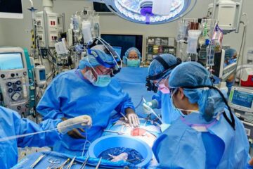
In the latest research, Henry Ford surgeons used robotic techniques to operate on seven kidney cancer patients between April and September 2012. In each case, they performed a partial nephrectomy, in which just the cancerous portion of the kidney is removed.
“What we've done is utilized a special type of device called a GelPoint trocar, that makes it easier to pass large things in and out of the abdomen through small incisions during minimally invasive surgery,” says Dr. Rogers.
“Through the gel point, we take a syringe that's been modified so we can pack and deliver ice through the body to the kidney. So when we clamp the blood supply to the kidney, it's packed in ice just as it would be in an open surgery.
“Once the tumor is removed, instead of setting it aside in the body so you can sew up the kidney, we can remove the tumor as soon as it's excised through the gel point, look at it and decide if we're happy with what's been removed. If there's any doubt, I can go right back in and cut more out.”
Dr. Rogers wrote in the study that others have tried various techniques to cool the kidney in minimally invasive surgery, but they “require specific equipment or expertise and are too complex or impractical for routine use.”
“Unfortunately, the majority of people today diagnosed with kidney cancer get their entire kidney removed,” Dr. Rogers says. “Not only that, they're getting it removed through an open approach, though a large incision that often requires removal a rib, when there are minimally invasive approaches, such as robotic surgery, available.”
One of the reasons more patients aren't given the option of a partial nephrectomy is because for the surgeon, it's technically challenging to do and much more difficult to perform than taking the entire kidney out.
Dr. Rogers explains: “In order to safely perform a partial nephrectomy, surgeons often have to clamp off the blood supply to the kidney to allow them to see the tumor and cut it out in a bloodless field. But once the blood supply is cut off to the kidney, there's only about 30 minutes before the kidney can experience irreversible damage. That means the surgeon has to be very technically skilled to remove the tumor, and sew the kidney back together in a very short time.
“Time is a barrier for many surgeons to offer the partial nephrectomy procedure to their patients. Or, for those who do, they'll offer the open approach to a partial nephrectomy, which means a bigger operation for the patient. There are two things surgeons who perform open nephrectomies have been able to claim, up until now, that they could do which minimally invasive surgeons could not.
First, when the surgeon is holding the kidney in the open approach and the clamp goes on the kidney to stop the blood flow, he can pack the kidney on ice to cool it, called renal hypothermia. It allows the surgery to extend the window of time he has to work without kidney damage,” says Dr. Rogers.
The other claim is that when the tumor is cut out, the surgeon can hold it and analyze it, to make sure that he is happy with what's been removed before sewing the kidney back up.
“Before, with a minimally invasive partial nephrectomy, it was very hard to get ice in through the small incision and to get the ice to stay where you want it. And then once the tumor's cut out, you really can't take it out of the body right away and look at it,” says Dr. Rogers.
“The reason why I'm so excited about what we've discovered and innovated at Henry Ford is that it cuts to the core of the real problem. We can now offer more patients partial nephrectomy through a minimally invasive approach. So any technology that allows patients to get a better surgery and a better outcome in a less invasive way, is going to be something that will benefit everyone.”
Note: You may request a pdf of the study or go online at http://www.sciencedirect.com/science/article/pii/S0302283812013607.
Media Contact
More Information:
http://www.hfhs.orgAll latest news from the category: Medical Engineering
The development of medical equipment, products and technical procedures is characterized by high research and development costs in a variety of fields related to the study of human medicine.
innovations-report provides informative and stimulating reports and articles on topics ranging from imaging processes, cell and tissue techniques, optical techniques, implants, orthopedic aids, clinical and medical office equipment, dialysis systems and x-ray/radiation monitoring devices to endoscopy, ultrasound, surgical techniques, and dental materials.
Newest articles

High-energy-density aqueous battery based on halogen multi-electron transfer
Traditional non-aqueous lithium-ion batteries have a high energy density, but their safety is compromised due to the flammable organic electrolytes they utilize. Aqueous batteries use water as the solvent for…

First-ever combined heart pump and pig kidney transplant
…gives new hope to patient with terminal illness. Surgeons at NYU Langone Health performed the first-ever combined mechanical heart pump and gene-edited pig kidney transplant surgery in a 54-year-old woman…

Biophysics: Testing how well biomarkers work
LMU researchers have developed a method to determine how reliably target proteins can be labeled using super-resolution fluorescence microscopy. Modern microscopy techniques make it possible to examine the inner workings…


