SNM 2010 Image of the Year: Molecular Imaging Shows Parathyroid Disease in Greater Detail
SNM’s 2010 Image of the Year illustrates the potential of hybrid molecular imaging to provide precise information about the location and function of a condition known as “hyperparathyroidism.” Researchers selected this image from more than 1,500 studies presented over the course of four days during SNM’s 57th Annual Meeting in Salt Lake City.
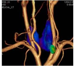
“Each year, SNM chooses an image that exemplifies the most cutting-edge molecular imaging research today, as well as illustrates the potential of molecular imaging to provide physicians with a critical component for the detection and diagnosis of disease,” said Michael M. Graham, Ph.D., M.D., immediate past-president of SNM. “This year’s Image of the Year provides an example of a novel imaging presentation, using a combination of single photon emission computed tomography (SPECT) with high resolution CT angiography, which pinpoints the abnormally functioning parathyroid adenoma and the arteries feeding it. With this information, physicians may be able determine the exact location and size of the abnormal gland and plan minimally invasive surgery that reduces operative time, thus improving patient care.”
The SNM Image of the Year shows the potential of fusion of high-resolution 3D anatomy with functional SPECT images to provide critical information to help physicians to diagnose and treat hyperparathyroidism, an endocrine disease that occurs when the parathyroid glands develop a small adenoma, a benign tumor that produces too much hormone and causes high levels of calcium in the blood. It is usually treated by invasive, exploratory surgery. Using fusion images, physicians can obtain detailed information about the anatomical localization, blood supply and metabolism of the overactive parathyroid adenoma.
In this study, researchers scanned 31 patients with symptoms of primary hyperparathyroidism using a nuclear medicine technique called MIBI, combined with SPECT and multidetector computed tomography (MDCT). Researchers obtained thin-slice multiplanar reconstruction images of the neck using a 64-row MDCT with contrast enhancement. When the enlarged gland was successfully identified, volume-rendered images of the thyroid and parathyroid with feeding arteries were generated. Then, 2- and 3-dimensional fusion images were also obtained using dedicated workstations. The diagnostic value of 3-dimensional SPECT/CT fusion images was compared with those by MIBI SPECT alone and by ultrasound. The study shows that the hybrid molecular imaging technique was more effective than single modality scanning alone.
A total of 34 glands were identified by surgery. SPECT/CT fusion image, MIBI SPECT and ultrasound identified 32 (94%), 27 (79%) and 27 (79%) adenomas, respectively. The fusion imaging technique identified five glands that were missed by ultrasound and MIBI SPECT. The fusion images successfully showed feeding arteries in 29 adenomas. With the use of fusion images for navigation, preliminary results in eight patients showed that operation time is decreased by approximately 82% compared to studies performed without fusion images.
According to the National Institutes of Health, approximately 100,000 Americans develop hyperparathyroidism each year. Women outnumber men two to one, and risk increases with age. In women 60 years and older, two out of 1,000 will develop hyperparathyroidism each year.
Abstract 200: K. Nakada, I. Sakuma, M. Sakurai, K. Noriyasu, Hokko Memorial Hospital, Sapporo, Japan; N. Takada, Kaisei Hospital, Sapporo, Japan; H. Takahashi, Hokkaido University Hospital, Sapporo, Japan. “Clinical Value of Fusion Images of MIBI SPECT and Enhanced MDCT Registered by Workstation in Primary Hyperparathyroidism.” SNM’s 57th Annual Meeting, June 5–9, 2010, Salt Lake City, Utah.
About SNM—Advancing Molecular Imaging and Therapy
SNM is an international scientific and medical organization dedicated to raising public awareness about what molecular imaging is and how it can help provide patients with the best health care possible. SNM members specialize in molecular imaging, a vital element of today’s medical practice that adds an additional dimension to diagnosis, changing the way common and devastating diseases are understood and treated.
SNM’s more than 17,000 members set the standard for molecular imaging and nuclear medicine practice by creating guidelines, sharing information through journals and meetings and leading advocacy on key issues that affect molecular imaging and therapy research and practice. For more information, visit http://www.snm.org.
Media Contact
More Information:
http://www.snm.orgAll latest news from the category: Medical Engineering
The development of medical equipment, products and technical procedures is characterized by high research and development costs in a variety of fields related to the study of human medicine.
innovations-report provides informative and stimulating reports and articles on topics ranging from imaging processes, cell and tissue techniques, optical techniques, implants, orthopedic aids, clinical and medical office equipment, dialysis systems and x-ray/radiation monitoring devices to endoscopy, ultrasound, surgical techniques, and dental materials.


Newest articles

High-energy-density aqueous battery based on halogen multi-electron transfer
Traditional non-aqueous lithium-ion batteries have a high energy density, but their safety is compromised due to the flammable organic electrolytes they utilize. Aqueous batteries use water as the solvent for…

First-ever combined heart pump and pig kidney transplant
…gives new hope to patient with terminal illness. Surgeons at NYU Langone Health performed the first-ever combined mechanical heart pump and gene-edited pig kidney transplant surgery in a 54-year-old woman…

Biophysics: Testing how well biomarkers work
LMU researchers have developed a method to determine how reliably target proteins can be labeled using super-resolution fluorescence microscopy. Modern microscopy techniques make it possible to examine the inner workings…

