New brain scan technology could save babies’ lives
scanning made simple, the new technology is designed to cause minimal disturbance to the baby
A revolutionary portable brain scanner under development could aid the treatment, and in some cases help save the lives, of premature and newborn babies in intensive care.
By providing vital information about brain function at the cot side, the scanner avoids the need to move critically ill babies to conventional scanning facilities, which may involve sedating them and has a degree of risk. The data produced by the new scanner can be used to diagnose and assess conditions such as brain haemorrhages and brain damage, and to inform decisions on effective treatment options.
A prototype of the scanner, called MONSTIR, has been developed by researchers at UCL (University College London) with funding from the Engineering and Physical Sciences Research Council (EPSRC) and the Wellcome Trust. Now, also with EPSRC funding, the team are aiming to reduce the size of the scanner and improve its speed of operation.
Currently, there are two main ways of performing brain scans on small babies. Magnetic resonance imaging (MRI) can provide data on brain function, but MRI scanners are large and static, and the baby may need to be sedated and wheeled to the scanner for the procedure to be carried out. The other alternative, ultrasound, can be performed at the cot side and is effective at revealing brain anatomy, but cannot show how the brain is actually functioning, e.g. in terms of oxygen supply and blood flow.
Combining the advantages of MRI and ultrasound but avoiding their disadvantages, MONSTIR, the first scanner of its kind in the world, uses an innovative technique called optical tomography to generate images showing how the brain is working. In optical tomography, light passes through body tissue and is then analysed by computer to provide information about the tissue.
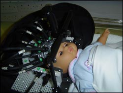
A helmet incorporating 32 light detectors and 32 sources of completely safe, low-intensity laser light is placed on the baby’s head. The sources produce short flashes and the detectors measure the amount of light that reaches them through the brain and the time the light takes to travel. A software package also developed with EPSRC funding uses this information to build up a 3D image. This can show which parts of the brain are receiving oxygen, where blood is situated, evidence of brain damage etc.
MONSTIR is the size of a fridge-freezer and takes around 8 minutes to generate an image. The aim is to produce a version that is half this size, 5 times faster, even more accurate and geared for clinical use. The potential use of the technology in breast imaging to aid cancer prevention and treatment is also being trialled.
Dr Adam Gibson is a key member of the multi-disciplinary MONSTIR team at UCL that includes medics, physiologists and mathematicians. He says: “The technology we’re developing has the potential to produce high-quality images at the cot side and is also cheaper than MRI. It could make an important contribution to the care and treatment of critically ill babies. Scanners could be available commercially within a few years.”
Media Contact
More Information:
http://www.epsrc.ac.ukAll latest news from the category: Medical Engineering
The development of medical equipment, products and technical procedures is characterized by high research and development costs in a variety of fields related to the study of human medicine.
innovations-report provides informative and stimulating reports and articles on topics ranging from imaging processes, cell and tissue techniques, optical techniques, implants, orthopedic aids, clinical and medical office equipment, dialysis systems and x-ray/radiation monitoring devices to endoscopy, ultrasound, surgical techniques, and dental materials.


Newest articles

Silicon Carbide Innovation Alliance to drive industrial-scale semiconductor work
Known for its ability to withstand extreme environments and high voltages, silicon carbide (SiC) is a semiconducting material made up of silicon and carbon atoms arranged into crystals that is…

New SPECT/CT technique shows impressive biomarker identification
…offers increased access for prostate cancer patients. A novel SPECT/CT acquisition method can accurately detect radiopharmaceutical biodistribution in a convenient manner for prostate cancer patients, opening the door for more…

How 3D printers can give robots a soft touch
Soft skin coverings and touch sensors have emerged as a promising feature for robots that are both safer and more intuitive for human interaction, but they are expensive and difficult…

