Insights into Micromillimeters
“TIGA,” the new high-tech imaging center at the University of Heidelberg founded in cooperation with the Japanese company Hamamatsu, provides deep insights: a high-tech robot makes it possible for the first time to automatically reproduce and evaluate tissue slices only micromillimeters thick – an important aid for researchers in understanding cancer or in following in detail the effect of treatment on cells and tissue.
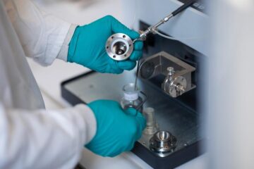
The Hamamatsu Tissue Imaging and Analysis (TIGA) Center is a cooperative effort between the Institutes of Pathology and of Medical Biometry and Informatics at the University of Heidelberg and the Japanese company Hamamatsu Photonics. In addition, it belongs to BIOQUANT, the research center for quantitative biology at the University of Heidelberg. At its core is the imaging robot “NanoZoomer” from Hamamatsu Photonics: the robot scans the tissue slices and displays them on the monitor for researchers at ultra high resolution and in various planes.
“Technically, this has brought the fully automatic evaluation of tissue changes and approaches for new therapy within our grasp,” states Professor Dr. Peter Schirmacher, Director of the Institute for Pathology at Heidelberg University Hospital. This would represent a new milestone in pathology.
Detailed images help understand diseases
Which proteins are formed to a greater degree in cancer cells? How is tumor tissue changed during radiation treatment? Thanks to the NanoZoomer’s high-resolution images and special evaluation programs, researchers in the future will be able to evaluate tissue and cell samples more quickly and accurately and gain important new insights for therapy tailored to the individual patient, for example for breast cancer.
In the future, the robot will be able to determine changes in cells and tissue fully automatically. “The NanoZoomer represents a quantum leap in tissue research,” says Dr. Niels Grabe of the Institute for Medical Biometry and Informatics and research director at the TIGA Center.
Virtual Tissue is modeled from data
The medical IT specialists use the NanoZoomer to evaluate huge quantities of data from tissues for their research. For example, Dr. Niels Grabe and his team used data to model virtual skin tissue. “On a computer model of human skin tissue we can test whether certain substances are toxic, for example,“ explains Dr. Grabe. “In the future, this could make it easier to develop potential new drugs.”
Hamamatsu recognized the many possible applications early on, so that new technological markets have now been opened up for them. “We are happy to have found two partners in the Heidelberg Institute of Pathology and the Institute of Medical Biometry and Informatics with whom we can develop concrete clinical uses and new applications for research,” said Hideo Hiruma, Managing Director of Hamamatsu Photonics, Japan.
Contact:
Dr. Niels Grabe
Research Director at the TIGA center
Tel.: +49 6221 / 56 5143
E-Mail: niels.grabe@med.uni-heidelberg.de
Professor Dr. Peter Schirmacher
Director of the Institute for Pathology
at Heidelberg University Hospital
Phone: +49 6221 / 56 2601
E-Mail: peter.schirmacher@med.uni-heidelberg.de
Hamamatsu Photonics, Germany and Japan:
Hamamatsu Germany is the German subsidiary of Hamamatsu Photonics K.K. (Japan), a leading manufacturer of devices for the generation and measurement of infrared, visible, and ultraviolet light. These devices include photodiodes, photomultiplier tubes, scientific light sources, infrared detectors, photoconductive cells, image sensors and integrated measurement systems for science and industry. The parent company is dedicated to the advancement of photonics through extensive research. This corporate philosophy results in state-of-the-art products which are used throughout the world in scientific, industrial, and commercial applications.
Institute of Pathology, University Heidelberg:
The Institute of Pathology at the University Heidelberg contributes to patient care, teaching, advanced training, quality management and research. Key task is the diagnostic evaluation of tissues (histology) and cell preparations (cytology). The Institute analyses more than 60.000 samples from operative and conservative medicine which are an elementary component of clinical diagnostics and therapy planning. The Institute is consulting in many areas, for example tumor diagnostics.
Institute of Medical Biometry and Informatics, University Heidelberg:
The Institute of Medical Biometry and Informatics at the University Heidelberg contributes to teaching, advanced training and clinical research. Biometry is concerned with the methodology and realization of therapeutic-, diagnostic- and meta studies. Research subjects of medical informatics includes bioinformatics/systems biology, knowledge based diagnosis and therapy, the management of health data, as well as medical image processing and pattern recognition. In collaboration with the University Heilbronn, the institute is conducting Germany’s eldest curriculum on medical informatics.
Requests by journalists:
Dr. Annette Tuffs
Head of Public Relations and Press Department
University Hospital of Heidelberg and
Medical Faculty of Heidelberg
Im Neuenheimer Feld 672
D-69120 Heidelberg
Germany
phone: +49 6221 / 56 45 36
fax: +49 6221 / 56 45 44
e-mail: annette.tuffs(at)med.uni-heidelberg.de
Media Contact
More Information:
http://www.med.uni-heidelberg.deAll latest news from the category: Medical Engineering
The development of medical equipment, products and technical procedures is characterized by high research and development costs in a variety of fields related to the study of human medicine.
innovations-report provides informative and stimulating reports and articles on topics ranging from imaging processes, cell and tissue techniques, optical techniques, implants, orthopedic aids, clinical and medical office equipment, dialysis systems and x-ray/radiation monitoring devices to endoscopy, ultrasound, surgical techniques, and dental materials.


Newest articles

Security vulnerability in browser interface
… allows computer access via graphics card. Researchers at Graz University of Technology were successful with three different side-channel attacks on graphics cards via the WebGPU browser interface. The attacks…
A closer look at mechanochemistry
Ferdi Schüth and his team at the Max Planck Institut für Kohlenforschung in Mülheim/Germany have been studying the phenomena of mechanochemistry for several years. But what actually happens at the…

Severe Vulnerabilities Discovered in Software to Protect Internet Routing
A research team from the National Research Center for Applied Cybersecurity ATHENE led by Prof. Dr. Haya Schulmann has uncovered 18 vulnerabilities in crucial software components of Resource Public Key…

