Novel rehabilitation device improves motor skills after stroke
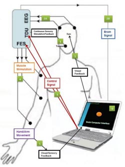
This is a schematic representation of the BCI-FES device indicating input and feedback components.<br><br>Credit: Radiological Society of North America<br>
“Each year, nearly 800,000 people suffer a new or recurrent stroke in the United States, and 50 percent of those have some degree of upper extremity disability,” said Vivek Prabhakaran, M.D., Ph.D., director of functional neuroimaging in radiology at the University of Wisconsin-Madison. “Rehabilitation sessions with our device allow patients to achieve an additional level of recovery and a higher quality of life.”
Dr. Prabhakaran, along with co-principal investigator Justin Williams, Ph.D., and a multidisciplinary team, built the new rehabilitation device by pairing a functional electrical stimulation (FES) system, which is currently used to help stroke patients recover limb function, and a brain control interface (BCI), which provides a direct communication pathway between the brain and this peripheral stimulation device.
In an FES system, electrical currents are used to activate nerves in paralyzed extremities. Using a computer and an electrode cap placed on the head, the new BCI-FES device (called the Closed-Loop Neural Activity-Triggered Stroke Rehabilitation Device) interprets electrical impulses from the brain and transmits the information to the FES.
“FES is a passive technique in that the electrical impulses move the patients' extremities for them,” Dr. Prabhakaran said. “When a patient using our device is asked to imagine or attempt to move his or her hand, the BCI translates that brain activity to a signal that triggers the FES. Our system adds an active component to the rehabilitation by linking brain activity to the peripheral stimulation device, which gives the patients direct control over their movement.”
The Wisconsin team conducted a small clinical trial of their rehabilitation device, enlisting eight patients with one hand affected by stroke. The patients were also able to serve as a control group by using their normal, unaffected hand. Patients in the study represented a wide range of stroke severity and amount of time elapsed since the stroke occurred. Despite having received standard rehabilitative care, the patients had varying degrees of residual motor deficits in their upper extremities. Each underwent nine to 15 rehabilitation sessions of two to three hours with the new device over a period of three to six weeks.
The patients also underwent functional magnetic resonance imaging (fMRI) and diffusion tensor imaging (DTI) before, at the mid-point of, at the end of, and one month following the rehabilitation period. fMRI is able to show which areas of the brain are activated while the patient performs a task, and DTI reveals the integrity of fibers within the white matter that connects the brain's functional areas.
Patients who suffered a stroke of moderate severity realized the greatest improvements to motor function following the rehabilitation sessions. Patients diagnosed with mild and severe strokes reported improved ability to complete activities of daily living following rehabilitation.
Dr. Prabhakaran said the results captured throughout the rehabilitation process—specifically the ratio of hemispheric involvement of motor areas—related well to the behavioral changes observed in patients. A comparison of pre-rehabilitation and post-rehabilitation fMRI results revealed reorganization in the regions of the brain responsible for motor function. DTI results over the course of the rehabilitation period revealed a gradual strengthening of the integrity of the fiber tracts.
“Our hope is that this device not only shortens rehabilitation time for stroke patients, but also that it brings a higher level of recovery than is achievable with the current standard of care,” Dr. Prabhakaran said. “We believe brain imaging will be helpful in both planning and tracking a stroke patient's therapy, as well as learning more about neuroplastic changes during recovery.”
Other co-authors are Dorothy Farrar-Edwards, Ph.D., Justin Sattin, M.D., Mitch Tyler, Ph.D., Veena A. Nair, Ph.D., Svyatoslav Vergun, B.S., Leo Walton, B.S., Jie Song, M.S., and Brittany Young, B.A., B.S.
Note: Copies of RSNA 2013 news releases and electronic images will be available online at RSNA.org/press13 beginning Monday, Dec. 2.
RSNA is an association of more than 53,000 radiologists, radiation oncologists, medical physicists and related scientists, promoting excellence in patient care and health care delivery through education, research and technologic innovation. The Society is based in Oak Brook, Ill. (RSNA.org)
For patient-friendly information on MRI, visit RadiologyInfo.org.
Media Contact
More Information:
http://www.rsna.orgAll latest news from the category: Medical Engineering
The development of medical equipment, products and technical procedures is characterized by high research and development costs in a variety of fields related to the study of human medicine.
innovations-report provides informative and stimulating reports and articles on topics ranging from imaging processes, cell and tissue techniques, optical techniques, implants, orthopedic aids, clinical and medical office equipment, dialysis systems and x-ray/radiation monitoring devices to endoscopy, ultrasound, surgical techniques, and dental materials.


Newest articles

Silicon Carbide Innovation Alliance to drive industrial-scale semiconductor work
Known for its ability to withstand extreme environments and high voltages, silicon carbide (SiC) is a semiconducting material made up of silicon and carbon atoms arranged into crystals that is…

New SPECT/CT technique shows impressive biomarker identification
…offers increased access for prostate cancer patients. A novel SPECT/CT acquisition method can accurately detect radiopharmaceutical biodistribution in a convenient manner for prostate cancer patients, opening the door for more…

How 3D printers can give robots a soft touch
Soft skin coverings and touch sensors have emerged as a promising feature for robots that are both safer and more intuitive for human interaction, but they are expensive and difficult…

