New PET radiotracer identifies inflammation in life-threatening atherosclerosis
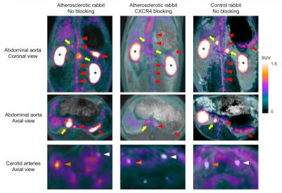
Note the high uptake of Ga-68-pentixafor on multi-planar reconstructions in the organs expressing CXCR4 such as the spleen (red arrows) and adrenal glands (yellow arrows), which was nearly completely blocked by the pre-injection of AMD 3100, a potent CXCR4 inhibitor. Strong accumulation of Ga-68-pentixafor was also found in the kidneys (asterisks) reflecting the renal clearance of the tracer. In addition, high, focal activities were detected in the abdominal aorta (red arrowheads) and right carotid artery (orange arrowheads) of atherosclerotic rabbits, whereas no significant signal could be detected in the non-injured left carotid artery (white arrowheads) of atherosclerotic and control rabbits, as well as in the abdominal aorta and right carotid artery of control rabbits. Furthermore, focal activities detected with PET in atherosclerotic plaques of the abdominal aorta and the right carotid artery decreased significantly when the same rabbit was re-imaged after blocking CXCR4 receptors. Credit: Fabien Hyafil,MD, PhD, Department of Nuclear Medicine, Klinikum Rechts der Isar, Technische Universität München, Munich, Germany
Atherosclerosis represents the main cause of heart attack and stroke. According to the Centers for Disease Control and Prevention, every year about 735,000 Americans have a heart attack and 800,000 have a stroke. Stroke kills more than 130,00 Americans a year, and about 610,000 die from cardiovascular disease.
Atherosclerosis develops over decades with the progressive accumulation of lipids, inflammatory cells and connective tissue within the inner layer of arterial walls leading to a local thickening of the vascular wall called atherosclerotic plaque.
These plaques can remain asymptomatic for years, but an inflammatory reaction can develop causing the plaques to rupture and stimulate clot formation. If a clot completely blocks an artery, no oxygen can reach the downstream tissue–resulting in the sudden development of heart attack or stroke. The challenge is to identify patients with these dangerous atherosclerotic plaques before a heart attack or stroke occurs.
Currently, there is no clinically available non-invasive imaging technique specifically to detect inflammation. F-18-fluorodeoxyglucose (FDG)-PET is being used but has important limitations. It is taken up by many cells other than inflammatory cells, including cardiac and brain cells.
The strong signal present in the organs next to the arteries limits the precise analysis of the radiotracer uptake in atherosclerotic plaques. In addition, patients need to fast at least six hours before FDG injection to avoid interferences with blood sugar and muscular uptake of the tracer that impair image quality.
“Ga-68-pentixafor binds more specifically to inflammatory cells than FDG and does not require the patient to fast for six hours before imaging,” explains Fabien Hyafil, MD, PhD, of Klinikum Rechts der Isar, Munich, Germany, and Bichat University Hospital, Assistance Publique, Hôpitaux de Paris, Paris, France.
In the study, the specific binding of Ga-68-pentixafor to inflammatory cells located in atherosclerotic plaques was first validated in an animal model. Seven atherosclerotic rabbits and five controls were imaged on a PET-MRI system after injection of the tracer. Resulting images clearly showed inflammation in plaques in the abdominal aorta and right carotid artery of the atherosclerotic rabbits. The researchers also confirmed with a small number of human patients that the radiotracer detected atherosclerotic plaques located in their carotid arteries.
Hyafil emphasizes, “This new radiotracer will strongly facilitate the imaging of inflammation in atherosclerotic plaques with PET and hopefully support the early detection and treatment of atherosclerosis, thus preventing heart attack or stroke.”
###
Authors of the article “Imaging the cytokine receptor CXCR4 in atherosclerotic plaques with the radiotracer 68Ga-pentixafor for positron emission tomography” include Fabien Hyafil, Klinikum Rechts der Isar, Munich, Germany, and Bichat University Hospital, Assistance Publique, Hôpitaux de Paris, Paris, France; Jaroslav Pelisek, Iina Laitinen, Miriam Mohring; Michael Kallmayer, Johannes Fischer, Christine Baumgartner, and Hans-Henning Eckstein, of Klinikum Rechts der Isar; Margret Schottelius, Katja Steiger, Andreas Poschenrieder, Johannes Notni, and Hans-Jürgen Wester, Technische Universität München, Garching, Germany; Yvonne Döring and Emiel P.C. van der Vorst, Ludwig-Maximilians-Universität München, Munich, Germany; Christian Weber, Ludwig-Maximilians-Universität München, Technische Universität München, Klinikum Rechts der Isar, DZHK partner site Munich Heart Alliance, and Maastricht University, The Netherlands; Christoph Rischpler, Stephan G. Nekolla, and Markus Schwaiger, Klinikum Rechts der Isar, Bichat University Hospital, Assistance Publique – Hôpitaux de Paris, Technische Universität München, Ludwig-Maximilians-Universität München, DZHK partner site Munich Heart Alliance.
This work was supported by the European Research Council Executive Agency through a Multimodal Molecular Imaging Advanced Research Grant (Grant number 294582), the Deutsche Forschungsgemeinschaft (SFB 824-B5 and SFB 1123-A1), and Deutsches Zentrum für Herz-Kreislauf Forschung through a high-risk, high-volume grant.
Please visit the SNMMI Media Center to view the PDF of the study, including images, and more information about molecular imaging and personalized medicine. To schedule an interview with the researchers, please contact Laurie Callahan at (703) 652-6773 or lcallahan@snmmi.org. Current and past issues of The Journal of Nuclear Medicine can be found online at http://jnm.
About the Society of Nuclear Medicine and Molecular Imaging
The Society of Nuclear Medicine and Molecular Imaging (SNMMI) is an international scientific and medical organization dedicated to raising public awareness about nuclear medicine and molecular imaging, a vital element of today's medical practice that adds an additional dimension to diagnosis, changing the way common and devastating diseases are understood and treated and helping provide patients with the best health care possible.
SNMMI's more than 17,000 members set the standard for molecular imaging and nuclear medicine practice by creating guidelines, sharing information through journals and meetings and leading advocacy on key issues that affect molecular imaging and therapy research and practice. For more information, visit http://www.
Media Contact
All latest news from the category: Medical Engineering
The development of medical equipment, products and technical procedures is characterized by high research and development costs in a variety of fields related to the study of human medicine.
innovations-report provides informative and stimulating reports and articles on topics ranging from imaging processes, cell and tissue techniques, optical techniques, implants, orthopedic aids, clinical and medical office equipment, dialysis systems and x-ray/radiation monitoring devices to endoscopy, ultrasound, surgical techniques, and dental materials.


Newest articles

Properties of new materials for microchips
… can now be measured well. Reseachers of Delft University of Technology demonstrated measuring performance properties of ultrathin silicon membranes. Making ever smaller and more powerful chips requires new ultrathin…

Floating solar’s potential
… to support sustainable development by addressing climate, water, and energy goals holistically. A new study published this week in Nature Energy raises the potential for floating solar photovoltaics (FPV)…

Skyrmions move at record speeds
… a step towards the computing of the future. An international research team led by scientists from the CNRS1 has discovered that the magnetic nanobubbles2 known as skyrmions can be…

