New insight into the brain’s hidden depths: Jena scientists develop minimally-invasive endoscope
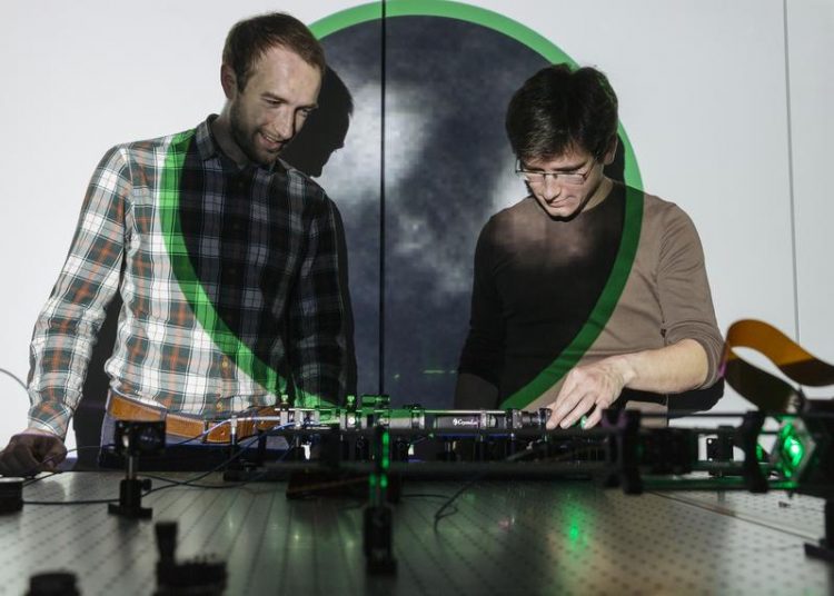
Sergey Turtaev (on the left) and Ivo T. Leite from Leibniz-IPHT. The projection behind them shows an image of neurons obtained deep inside the brain via a single multimode fibre. Sven Döring/ Agentur Focus
Using a hair-thin optical fibre, the researchers can look into deep brain areas of a living mouse as if through a keyhole. Recently introduced methods for holographic control of light propagation in complex media enable the use of a multimode fibre as an imaging tool.
Based on this new approach, the scientists designed a compact system for fluorescence imaging at the tip of a fibre, the most minimally invasive endoscopic probe reported thus far.
It offers a much smaller footprint as well as enhanced resolution compared to conventional endoscopes based on fibre bundles or graded-index lenses.
“We are very excited to see our technology making its first steps towards practical applications in neuroscience,” says Dr Sergey Turtaev from Leibniz-IPHT, lead author of the paper.
“For the first time, we have shown that it is possible to examine deep brain regions of a living animal model in a minimally invasive way and to achieve high-resolution images at the same time,” adds IPHT scientist Dr Ivo T. Leite.
Sergey and Ivo work in the research group led by IPHT scientist Prof. Tomáš Čižmár, who developed the holographic method for imaging through a single fibre. Using this approach, the research team succeeded in obtaining images of brain cells and neuronal processes in the visual cortex and hippocampus of living mice with resolution approaching one micrometre (i.e. one thousand times smaller than a millimetre).
Detailed observations within these areas are crucial for research into sensory perception, memory formation, and severe neuronal diseases such as Alzheimer’s.
Current investigation methods are strongly invasive, such that it is not possible to observe neuronal networks in these inner regions at work without massive destruction of the surrounding tissue – usual endoscopes comprised of hundreds of optical fibres are too large to penetrate such sensitive brain regions, while the neuronal structures are too tiny to be visualised by non-invasive imaging methods such as magnetic resonance imaging (MRI).
“This minimally invasive approach will enable neuroscientists to investigate functions of neurons in deep structures of the brain of behaving animals: without perturbing the neuronal circuits in action, it will be possible to reveal the activity of these neuronal circuits while the animal is exploring an environment or learning a new task,” explains project partner Dr Nathalie Rochefort from the University of Edinburgh.
Building up on this work, the research team now wants to address the current challenges of neuroscience, which will entail the delivery of advanced microscopy techniques through single fibre endoscopes. “Under the “Photonics for Life” flag of the Leibniz-IPHT and in the scope of the European Research Council funded project LIFEGATE, we will strive hard to prepare more significant advancements on this result, essentially funnelling the most advanced methods of modern microscopy deep inside the tissues of living and functioning organisms.” concludes Prof. Tomáš Čižmár.
Sergey Turtaev
sergey.turtaev(a)leibniz-ipht.de
+49 (0) 3641 · 206-225
Ivo Leite
ivo.leite(a)leibniz-ipht.de
+49 (0) 3641 · 206-225
https://www.nature.com/articles/s41377-018-0094-x
https://www.leibniz-ipht.de/en/institute/presse/news/detail/den-neuronen-bei-der…
Media Contact
All latest news from the category: Medical Engineering
The development of medical equipment, products and technical procedures is characterized by high research and development costs in a variety of fields related to the study of human medicine.
innovations-report provides informative and stimulating reports and articles on topics ranging from imaging processes, cell and tissue techniques, optical techniques, implants, orthopedic aids, clinical and medical office equipment, dialysis systems and x-ray/radiation monitoring devices to endoscopy, ultrasound, surgical techniques, and dental materials.


Newest articles

High-energy-density aqueous battery based on halogen multi-electron transfer
Traditional non-aqueous lithium-ion batteries have a high energy density, but their safety is compromised due to the flammable organic electrolytes they utilize. Aqueous batteries use water as the solvent for…

First-ever combined heart pump and pig kidney transplant
…gives new hope to patient with terminal illness. Surgeons at NYU Langone Health performed the first-ever combined mechanical heart pump and gene-edited pig kidney transplant surgery in a 54-year-old woman…

Biophysics: Testing how well biomarkers work
LMU researchers have developed a method to determine how reliably target proteins can be labeled using super-resolution fluorescence microscopy. Modern microscopy techniques make it possible to examine the inner workings…

