New diagnostic imaging techniques deemed safe in simulations
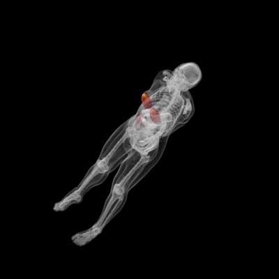
This shows simulated 3D dose measurements of the breast showing the dose imparted to the whole body. The dose is shown on a red and yellow color map, where yellow shows maximum dose. Credit: Duke Medicine
Using computer simulations, imaging the liver and breast with gamma or neutron radiation was found to be safe, delivering levels of radiation on par with conventional medical imaging, according to researchers at Duke Medicine.
The findings, published in the June issue of the journal Medical Physics, will help researchers to move testing of gamma and neutron imaging into animals and later humans.
Conventional medical imaging tools – including X-ray, ultrasound, CT and MRI – detect disease by finding the anatomy, or shape and size, of the abnormality. When using these tools to screen for cancer, a tumor must be large enough to be detected, and if found, a surgical biopsy is generally required to determine if it is benign or malignant.
Duke researchers are working to develop imaging technologies to detect disease in its earliest stages, much before the tumors grow large enough to be detected using conventional methods. Two imaging techniques they are researching are Neutron Stimulated Emission Computed Tomography and Gamma Stimulated Emission Computed Tomography.
Research has shown that many tumors have an out-of-balance concentration of trace-level elements naturally found in the body, such as aluminum and rubidium. These elements stray from their normal concentration levels at the earliest stages of tumor growth, potentially providing an early signal of disease.
The neutron and gamma imaging methods measure the concentrations of elements in the body, determining molecular properties without the need for a biopsy or injection of contrast media. The goal is for these tests to be able to distinguish between benign and malignant lesions, as well as healthy tissue.
“Gamma and neutron imaging may eventually be able to help us to detect cancer earlier without having to perform an invasive biopsy,” said Anuj Kapadia, Ph.D., assistant professor of radiology at Duke University School of Medicine and the study's senior author.
Gamma and neutron imaging may also have applications for patients undergoing cancer treatment. Patients currently wait weeks or months to see if their cancer is responding to a particular treatment and shrinking in size, but gamma and neutron imaging may be able to tell if a treatment is working earlier by detecting molecular changes directly within the tumor.
While improved diagnostic tests would provide clinicians with useful tools, one ongoing question is the safety of gamma and particularly neutron radiation. Upon entering the body, neutrons scatter considerably, with the possibility of reaching several vital organs. Thus, researchers have been concerned about how much radiation is absorbed in the targeted organ versus surrounding tissue. For instance, in a breast scan, how much radiation is delivered unnecessarily to the heart or lungs?
Using detailed computer simulations, Kapadia and his colleagues estimated the radiation dose delivered to the liver and breast using neutron and gamma imaging. They found that the majority of radiation was delivered to organs directly within the radiation beam, and a much lower dose was absorbed by tissue outside of the radiation beam.
In simulated breast scans, the radiation was almost entirely limited to the area of the breast being scanned. The dose to the breast accounted for 96 percent of the radiation in neutron scans and 99 percent in gamma scans. The heart and lungs received less than 1 percent of the radiation dose.
When imaging the liver in simulation, the neutron scan imparted the highest radiation dose to the liver, while in the gamma scan, the stomach wall absorbed the greatest amount of radiation given its location in the direct path of the beam. Further work is needed to reduce and better target gamma radiation doses in liver scans.
“The results show that despite the use of a highly scattering particle such as a neutron, the dose from neutron imaging is on par with other clinical imaging techniques such as X-ray CT,” Kapadia said. “Neutron and gamma radiation may become viable imaging alternatives if further testing proves them to be safe and effective.”
The researchers will use this information to move their studies into animals, and later, humans.
In addition to Kapadia, authors include Matthew D. Belley and William Paul Segars of Duke. The study was supported by the National Institutes of Health (R01-EB001838, T32-EB007185).
Media Contact
More Information:
http://www.duke.eduAll latest news from the category: Medical Engineering
The development of medical equipment, products and technical procedures is characterized by high research and development costs in a variety of fields related to the study of human medicine.
innovations-report provides informative and stimulating reports and articles on topics ranging from imaging processes, cell and tissue techniques, optical techniques, implants, orthopedic aids, clinical and medical office equipment, dialysis systems and x-ray/radiation monitoring devices to endoscopy, ultrasound, surgical techniques, and dental materials.


Newest articles

Security vulnerability in browser interface
… allows computer access via graphics card. Researchers at Graz University of Technology were successful with three different side-channel attacks on graphics cards via the WebGPU browser interface. The attacks…
A closer look at mechanochemistry
Ferdi Schüth and his team at the Max Planck Institut für Kohlenforschung in Mülheim/Germany have been studying the phenomena of mechanochemistry for several years. But what actually happens at the…

Severe Vulnerabilities Discovered in Software to Protect Internet Routing
A research team from the National Research Center for Applied Cybersecurity ATHENE led by Prof. Dr. Haya Schulmann has uncovered 18 vulnerabilities in crucial software components of Resource Public Key…

