Medica 2018: New software for a more efficient planning of minimally invasive surgery
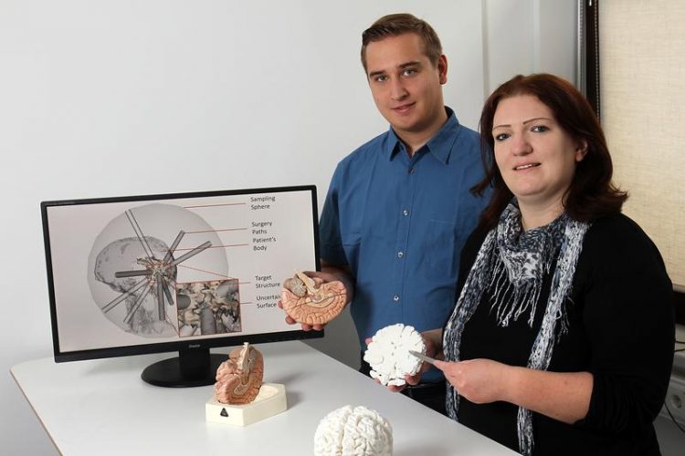
Christina Gillmann and Robin Maack are developing the software. Credits: Koziel/TUK
Many surgeons today use minimally invasive surgery to remove, for example, a tumour in the brain: A small hole in the cranium is sufficient for them to reach the affected area with a small probe. The probe is equipped with surgical instruments and a camera that provides the doctors with the necessary images.
“Such a surgery must be thoroughly planned beforehand,” says Dr Christina Gillmann of the Technische Universität Kaiserslautern (TUK). “Surgeons must locate the ideal route, i.e. the surgical channel, to the affected area, which damages as little important tissue in the brain as possible.”
For this purpose, they use images from magnetic resonance tomography (MRT) taken before the surgery. But other diagnostic procedures, such as computer tomography, are also used for such planning, depending on the disease in question.
These X-ray techniques provide physicians with images of patients on which they can see the body part to be operated in grey scales. “This data is often examined layer by layer in everyday clinical practice, however, with this technique it is often difficult to determine in which tissue layers there might be a suitable surgical channel,” explains the scientist.
A new procedure, on which Gillmann and her team are working, could remedy this problem: It enables doctors to plan their operations intuitively. “The computer program displays the individual tissue layers that are affected by a surgical channel,” says the computer scientist.
“This allows different channels to be compared and risks to be discussed.” In this way, doctors can also identify possible complications that might occur during the operation. “For example, the surgical team could discuss which route is the most appropriate for the individual patient,” Gillmann continues.
The computer scientists at the TUK use various medical images as a data basis for their method. Using their own calculation methods, they re-evaluate this image data. “We can visually separate and display the individual tissue layers so that it is easier to see where a surgical channel should be located,” explains Gillmann. The researchers have designed their technology in such a way that it is easy to use for surgeons.
The software is still in the development stage. “It will take a few more years before it can be used in clinical daily routine,” the computer scientist continues. The Kaiserslautern researchers are working closely with the Premier Health Hospital in Dayton, Ohio, USA.
Gillmann is researching in the field of “Computer Graphics and Human Computer Interaction” under Professor Dr Hans Hagen. Over the lst two decades, the research group has been working on processing medical imaging data in such a way that they can be used simply and reliably in everyday clinical practice. For example, they have succeeded in using their methods to separate tumours from healthy tissue more clearly in images. In their projects, the computer scientists work closely with various partners, including the University Hospital Leipzig and the Premier Health Hospital in Ohio.
Dr Christina Gillmann
Computer Graphics and Human Computer Interaction
Phone: +49(0)631205-2642
E-mail: c_gillma(at)cs.uni-kl.de
Media Contact
More Information:
http://www.uni-kl.deAll latest news from the category: Medical Engineering
The development of medical equipment, products and technical procedures is characterized by high research and development costs in a variety of fields related to the study of human medicine.
innovations-report provides informative and stimulating reports and articles on topics ranging from imaging processes, cell and tissue techniques, optical techniques, implants, orthopedic aids, clinical and medical office equipment, dialysis systems and x-ray/radiation monitoring devices to endoscopy, ultrasound, surgical techniques, and dental materials.


Newest articles

Silicon Carbide Innovation Alliance to drive industrial-scale semiconductor work
Known for its ability to withstand extreme environments and high voltages, silicon carbide (SiC) is a semiconducting material made up of silicon and carbon atoms arranged into crystals that is…

New SPECT/CT technique shows impressive biomarker identification
…offers increased access for prostate cancer patients. A novel SPECT/CT acquisition method can accurately detect radiopharmaceutical biodistribution in a convenient manner for prostate cancer patients, opening the door for more…

How 3D printers can give robots a soft touch
Soft skin coverings and touch sensors have emerged as a promising feature for robots that are both safer and more intuitive for human interaction, but they are expensive and difficult…

