Imaging technology allows visualization of nanoscale structures inside whole cells
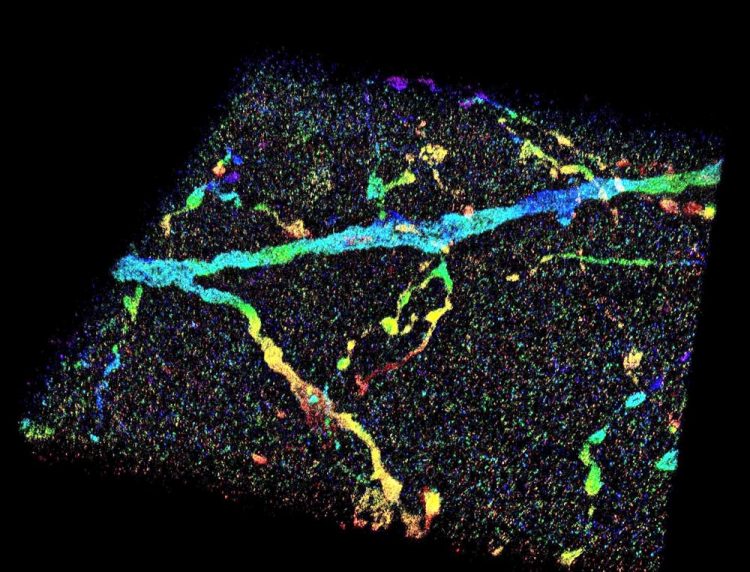
This image shows a 3D super-resolution reconstruction of dendrites in primary visual cortex. Purdue University innovators created an imaging tool that allows visualization of nanoscale structures inside whole cells and tissues. Credit: Fang Huang/Purdue University
Since Robert Hooke's first description of a cell in Micrographia 350 years ago, microscopy has played an important role in understanding the rules of life.
However, the smallest resolvable feature, the resolution, is restricted by the wave nature of light. This century-old barrier has restricted understanding of cellular functions, interactions and dynamics, particularly at the sub-micron to nanometer scale.
Super-resolution fluorescence microscopy overcomes this fundamental limit, offering up to tenfold improvement in resolution, and allows scientists to visualize the inner workings of cells and biomolecules at unprecedented spatial resolution.
Such resolving capability is impeded, however, when observing inside whole-cell or tissue specimens, such as the ones often analyzed during the studies of the cancer or the brain. Light signals, emitted from molecules inside a specimen, travel through different parts of cell or tissue structures at different speeds and result in aberrations, which will deteriorate the image.
Now, Purdue University researchers have developed a new technology to overcome this challenge.
“Our technology allows us to measure wavefront distortions induced by the specimen, either a cell or a tissue, directly from the signals generated by single molecules – tiny light sources attached to the cellular structures of interest,” said Fang Huang, an assistant professor of biomedical engineering in Purdue's College of Engineering. “By knowing the distortion induced, we can pinpoint the positions of individual molecules at high precision and accuracy. We obtain thousands to millions of coordinates of individual molecules within a cell or tissue volume and use these coordinates to reveal the nanoscale architectures of specimen constituents.”
The Purdue team's technology is recently published in Nature Methods. A video showing an animated 3D super-resolution is available at https:/
“During three-dimensional super-resolution imaging, we record thousands to millions of emission patterns of single fluorescent molecules,” said Fan Xu, a postdoctoral associate in Huang's lab and a co-first author of the publication. “These emission patterns can be regarded as random observations at various axial positions sampled from the underlying 3D point-spread function describing the shapes of these emission patterns at different depths, which we aim to retrieve. Our technology uses two steps: assignment and update, to iteratively retrieve the wavefront distortion and the 3D responses from the recorded single molecule dataset containing emission patterns of molecules at arbitrary locations.”
The Purdue technology allows finding the positions of biomolecules with a precision down to a few nanometers inside whole cells and tissues and therefore, resolving cellular and tissue architectures with high resolution and fidelity.
“This advancement expands the routine applicability of super-resolution microscopy from selected cellular targets near coverslips to intra- and extra-cellular targets deep inside tissues,” said Donghan Ma, a postdoctoral researcher in Huang's lab and a co-first author of the publication. “This newfound capacity of visualization could allow for better understanding for neurodegenerative diseases such as Alzheimer's, and many other diseases affecting the brain and various parts inside the body.”
The National Institutes of Health provided major support for the research.
Other members of the research team include Gary Landreth, a professor from Indiana University's School of Medicine; Sarah Calve, an associate professor of biomedical engineering in Purdue's College of Engineering (currently an associate professor of mechanical engineering at the University of Colorado Boulder); Peng Yin, a professor from Harvard Medical School; and Alexander Chubykin, an assistant professor of biological sciences at Purdue. The complete list of authors can be found in Nature Methods.
“This technical advancement is startling and will fundamentally change the precision with which we evaluate the pathological features of Alzheimer's disease,” Landreth said. “We are able to see smaller and smaller objects and their interactions with each other, which helps reveal structure complexities we have not appreciated before.”
Calve said the technology is a step forward in regenerative therapies to help promote repair within the body.
“This development is critical for understanding tissue biology and being able to visualize structural changes,” Calve said.
Chubykin, whose lab focuses on autism and diseases affecting the brain, said the high-resolution imaging technology provides a new method for understanding impairments in the brain.
“This is a tremendous breakthrough in terms of functional and structural analyses,” Chubykin said. “We can see a much more detailed view of the brain and even mark specific neurons with genetic tools for further study.”
The team worked with the Purdue Research Foundation Office of Technology Commercialization to patent the technology. The office recently moved into the Convergence Center for Innovation and Collaboration in Discovery Park District, adjacent to the Purdue campus.
###
The inventors are looking for partners to commercialize their technology. For more information on licensing this innovation, contact Dipak Narula of OTC at dnarula@prf.org.
About Purdue Research Foundation Office of Technology Commercialization
The Purdue Research Foundation Office of Technology Commercialization operates one of the most comprehensive technology transfer programs among leading research universities in the U.S. Services provided by this office support the economic development initiatives of Purdue University and benefit the university's academic activities through commercializing, licensing and protecting Purdue intellectual property. The office recently moved into the Convergence Center for Innovation and Collaboration in Discovery Park District, adjacent to the Purdue campus. In fiscal year 2019, the office reported 136 deals finalized with 231 technologies signed, 380 disclosures received and 141 issued U.S. patents. The office is managed by the Purdue Research Foundation, which received the 2019 Innovation and Economic Prosperity Universities Award for Place from the Association of Public and Land-grant Universities. In 2020, IPWatchdog Institute ranked Purdue third nationally in startup creation and in the top 20 for patents. The Purdue Research Foundation is a private, nonprofit foundation created to advance the mission of Purdue University. Contact otcip@prf.org for more information.
About Purdue University
Purdue University is a top public research institution developing practical solutions to today's toughest challenges. Ranked the No. 6 Most Innovative University in the United States by U.S. News & World Report, Purdue delivers world-changing research and out-of-this-world discovery. Committed to hands-on and online, real-world learning, Purdue offers a transformative education to all. Committed to affordability and accessibility, Purdue has frozen tuition and most fees at 2012-13 levels, enabling more students than ever to graduate debt-free. See how Purdue never stops in the persistent pursuit of the next giant leap at purdue.edu.
Writer: Chris Adam, 765-588-3341, cladam@prf.org
Sources: Fang Huang, fanghuang@purdue.edu
Fan Xu, xu240@purdue.edu
Donghan Ma, ma511@purdue.edu
Gary Landreth, glandret@iu.edu
Sarah Calve, Sarah.Calve@colorado.edu
Alex Chubykin, chubykin@purdue.edu
Media Contact
All latest news from the category: Medical Engineering
The development of medical equipment, products and technical procedures is characterized by high research and development costs in a variety of fields related to the study of human medicine.
innovations-report provides informative and stimulating reports and articles on topics ranging from imaging processes, cell and tissue techniques, optical techniques, implants, orthopedic aids, clinical and medical office equipment, dialysis systems and x-ray/radiation monitoring devices to endoscopy, ultrasound, surgical techniques, and dental materials.


Newest articles

Silicon Carbide Innovation Alliance to drive industrial-scale semiconductor work
Known for its ability to withstand extreme environments and high voltages, silicon carbide (SiC) is a semiconducting material made up of silicon and carbon atoms arranged into crystals that is…

New SPECT/CT technique shows impressive biomarker identification
…offers increased access for prostate cancer patients. A novel SPECT/CT acquisition method can accurately detect radiopharmaceutical biodistribution in a convenient manner for prostate cancer patients, opening the door for more…

How 3D printers can give robots a soft touch
Soft skin coverings and touch sensors have emerged as a promising feature for robots that are both safer and more intuitive for human interaction, but they are expensive and difficult…

