New imaging technique visualizes cancer during surgery
The technique has now been successfully tested on nine patients diagnosed with ovarian cancer. There are plans to apply this imaging concept also to minimally invasive and endoscopic procedures.
Ovarian cancer is one of the most frequent forms of cancer that affect women. As tumors can initially grow unchecked in the abdomen without causing any major symptoms, patients are usually diagnosed at an advanced stage and have to undergo surgery plus chemotherapy. During the operation, surgeons attempt to remove all tumor deposits as this leads to improved patient prognosis. To do this, however, they primarily have to rely on visual inspection and palpation – an enormous challenge especially in the case of small tumor nests or remaining tumor borders after the primary tumor excision.
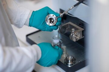
Yet surgeons could now be getting support from a new multispectral fluorescence imaging system developed by a team of researchers in Munich, headed by Vasilis Ntziachristos, Professor of Biological Imaging. A study carried out on nine patients with ovarian cancer has shown that the new system can be used to localize cancer cells during surgery. Before the operation, the patients were injected with folic acid chemically coupled to a green fluorescent dye. Most ovarian tumors have a protein molecule on their surface that bonds with folic acid and transports it inside the cell. This protein is known as the folate receptor alpha. During abdominal surgery, the surgeon can then shine a special laser light onto the patient’s ovaries, causing the green-labeled folic acid inside the cancer cells to emit light. Healthy tissue remains dark.
The fluorescent cancer cells, however, cannot be detected by the naked eye. Three cameras, mounted on a pivoting support arm over the operating table, detect optical and fluorescent signals at multiple spectral bands and then correct for light variations due to illumination and tissue discolorations in order to provide truly accurate fluorescence images that can be simultaneously displayed with corresponding color images on monitors in the operating room. The surgeon can thus check whether all the cancer cells have been removed by inspecting for remnant fluorescence light. In eight of the nine patients, doctors were able to remove small clusters of tumor cells that might otherwise have gone undetected. The multispectral fluorescence imaging system has thus passed its first OR test. However, it will have to prove its value to improve clinical outcome in further operations before it can be deployed for routine surgical procedures.
The researchers in Munich and Groningen also want to further develop the camera system so it can be used to detect other forms of tumors during operations. Of significant importance in future developments is the ability to offer accurate fluorescence imaging so that data collected reflect true presence of disease. “The use of advanced, real-time optical technology will allow us to standardize data collection and accuracy so that studies performed at multiple clinical centers can be accurately compared and analyzed” explains Prof. Vasilis Ntziachristos. This is important for the clinical acceptance of the technology and its approval by regulatory agencies. In the future patient selection through personalized medicine approaches, for example by obtaining a molecular profile of the tumor of each patient, would further enable custom-tailored surgical treatment of improved accuracy. The team is also planning to build a version for minimally invasive operations.
Acknowledgment: The folic acid chemically coupled to a green fluorescent dye was provided by Phil Low of Purdue University.
Publication:
Intraoperative tumor-specific fluorescence imaging in ovarian cancer by folate receptor-alpha targeting: first in-human results
Goolitzen M van Dam et al., Nature Medicine, Sept 2011, DOI: 10.1038/nm.2472
Contact:
Prof. Vasilis Ntziachristos
Institute for Biological Imaging
Technische Universität München / Helmholtz Zentrum München
Tel. +49 89 3187 4139
E-mail: v.ntziachristos[at]tum.de
Media Contact
More Information:
http://www.tum.deAll latest news from the category: Medical Engineering
The development of medical equipment, products and technical procedures is characterized by high research and development costs in a variety of fields related to the study of human medicine.
innovations-report provides informative and stimulating reports and articles on topics ranging from imaging processes, cell and tissue techniques, optical techniques, implants, orthopedic aids, clinical and medical office equipment, dialysis systems and x-ray/radiation monitoring devices to endoscopy, ultrasound, surgical techniques, and dental materials.


Newest articles

Security vulnerability in browser interface
… allows computer access via graphics card. Researchers at Graz University of Technology were successful with three different side-channel attacks on graphics cards via the WebGPU browser interface. The attacks…
A closer look at mechanochemistry
Ferdi Schüth and his team at the Max Planck Institut für Kohlenforschung in Mülheim/Germany have been studying the phenomena of mechanochemistry for several years. But what actually happens at the…

Severe Vulnerabilities Discovered in Software to Protect Internet Routing
A research team from the National Research Center for Applied Cybersecurity ATHENE led by Prof. Dr. Haya Schulmann has uncovered 18 vulnerabilities in crucial software components of Resource Public Key…

