New imaging tech promising for diagnosing cardiovascular disease, diabetes
The new method could be used to take precise three-dimensional images of plaques lining arteries, said Ji-Xin Cheng, an associate professor of biomedical engineering and chemistry at Purdue University.
Other imaging methods that provide molecular information are unable to penetrate tissue deep enough to reveal the three-dimensional structure of the plaques, but being able to do so would make better diagnoses possible, he said.
“You would have to cut a cross section of an artery to really see the three-dimensional structure of the plaque,” Cheng said. “Obviously, that can't be used for living patients.”
The imaging reveals the presence of carbon-hydrogen bonds making up lipid molecules in arterial plaques that cause heart disease. The method also might be used to detect fat molecules in muscles to diagnose diabetes and for other lipid-related disorders, including neurological conditions and brain trauma. The technique also reveals nitrogen-hydrogen bonds making up proteins, meaning the imaging tool also might be useful for diagnosing other diseases and to study collagen's role in scar formation.
“Being able to key on specific chemical bonds is expected to open a completely new direction for the field,” Cheng said
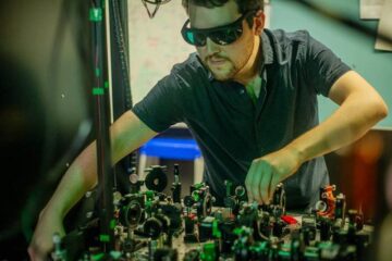
Findings are detailed in a paper to be published in Physical Review Letters and expected to appear in the June 17 issue. The findings represent the culmination of four years of research led by Cheng and doctoral student Han-Wei Wang.
The new technique uses nanosecond laser pulses in the near-infrared range of the spectrum. The laser generates molecular “overtone” vibrations, or wavelengths that are not absorbed by the blood. The pulsed laser causes tissue to heat and expand locally, generating pressure waves at the ultrasound frequency that can be picked up with a device called a transducer.
“We are working to miniaturize the system so that we can build an endoscope to put into blood vessels using a catheter,” Cheng said. “This would enable us to see the exact nature of plaque formation in the walls of arteries to better quantify and diagnose cardiovascular disease.”
Lihong Wang, Gene K. Beare Distinguished Professor of Biomedical Engineering at Washington University in St. Louis, is a pioneer of using the “photoacoustic” imaging of blood vessels based on the absorption of light by the electrons in hemoglobin.
The Purdue researchers are the first to show that a strong photoacoustic signal can arise from the absorption of light by the chemical bonds in molecules. The near-infrared laser causes enough heating to generate ultrasound but not enough to damage tissue.
“You can measure the time delay between the laser and the ultrasound waves, and this gives you a precise distance, which enables you to image layers of the tissues for three-dimensional pictures,” Cheng said. “You do one scan and get all the cross sections. Our initial target is cardiovascular disease, but there are other potential applications, including diabetes and neurological conditions.”
The approach represents a major improvement over another imaging technique, called coherent anti-Stokes Raman scattering, or CARS, which has been used by the Purdue-based lab to study three-dimensional plaque formation in arteries.
Also leading the research are Michael Sturek, chair of the Department of Cellular and Integrative Physiology at the Indiana University School of Medicine; Robert P. Lucht, Purdue's Ralph and Bettye Bailey Professor of Combustion in Mechanical Engineering; and David Umulis, a Purdue assistant professor of agricultural and biological engineering. Other authors of the paper include Purdue graduate students Ning Chai, Pu Wang and Wei Dou and Washington University postdoctoral researcher Song Hu.
Findings are based on research with pig tissues in laboratory samples and also with live fruit flies.
“You can see fat inside fly larvae, representing the potential to study how obesity affects physiology in humans,” Cheng said.
Research funding came from the National Institutes of Health and American Heart Association.
Writer: Emil Venere, 765-494-4709, venere@purdue.edu
Source: Ji-Xin Cheng, 765-494-4335, jcheng@purdue.edu
Note to Journalists: Ji-Xin Cheng is pronounced “Gee-Shin.” An electronic copy of the paper is available from Emil Venere, Purdue News Service, at 765-494-4709, venere@purdue.edu
Media Contact
More Information:
http://www.purdue.eduAll latest news from the category: Medical Engineering
The development of medical equipment, products and technical procedures is characterized by high research and development costs in a variety of fields related to the study of human medicine.
innovations-report provides informative and stimulating reports and articles on topics ranging from imaging processes, cell and tissue techniques, optical techniques, implants, orthopedic aids, clinical and medical office equipment, dialysis systems and x-ray/radiation monitoring devices to endoscopy, ultrasound, surgical techniques, and dental materials.


Newest articles
Combatting disruptive ‘noise’ in quantum communication
In a significant milestone for quantum communication technology, an experiment has demonstrated how networks can be leveraged to combat disruptive ‘noise’ in quantum communications. The international effort led by researchers…
Stretchable quantum dot display
Intrinsically stretchable quantum dot-based light-emitting diodes achieved record-breaking performance. A team of South Korean scientists led by Professor KIM Dae-Hyeong of the Center for Nanoparticle Research within the Institute for…
Internet can achieve quantum speed with light saved as sound
Researchers at the University of Copenhagen’s Niels Bohr Institute have developed a new way to create quantum memory: A small drum can store data sent with light in its sonic…

