Hybrid microscope could bring digital biopsy to the clinic
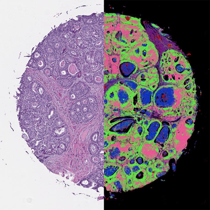
Machine-learning tools can analyze the data from the infrared-optical hybrid microscope to create digital versions of standard dyes, left, or to identify tissue types based on their chemical composition, right. Images courtesy of Rohit Bhargava, University of Illinois
Pairing infrared measurements with high-resolution optical images and machine learning algorithms, the researchers created digital biopsies that closely correlated with traditional pathology techniques and also outperformed state-of-the-art infrared microscopes.
Led by Rohit Bhargava, a professor of bioengineering and the director of the Cancer Center at Illinois, the group published its results in the Proceedings of the National Academy of Sciences.
“The advantage is that no stains are required, and both the organization of cells and their chemistry can be measured. Measuring the chemistry of tumor cells and their microenvironment can lead to better cancer diagnoses and better understanding of the disease,” Bhargava said.
The gold standard of tissue pathology is to add dyes or stains so that pathologists can see the shapes and patterns of the cells under a microscope. However, it can be difficult to distinguish cancer from healthy tissue or to pinpoint the boundaries of a tumor, and in many cases diagnosis is subjective.
“For more than a century, we have relied on adding dyes to human tissue biopsies to diagnose tumors. However, the shape and color induced by the dye provide very limited information about the underlying molecular changes that drive cancer,” Bhargava said.
Technologies like infrared microscopy can measure the molecular composition of tissue, providing quantitative measures that can distinguish cell types. Unfortunately, infrared microscopes are expensive and the samples require special preparation and handling, making them impractical for the vast majority of clinical and research settings.
Bhargava's group developed its hybrid microscope by adding an infrared laser and a specialized microscope lens, called an interference objective, to an optical camera. The infrared-optical hybrid measures both infrared data and a high-resolution optical image with a light microscope – the kind ubiquitous in clinics and labs.
“We built the hybrid microscope from off-the-shelf components. This is important because it allows others to easily build their own microscope or upgrade an existing microscope,” said Martin Schnell, a postdoctoral fellow in Bhargava's group and first author of the paper.
Combining the two techniques harnesses the strengths of both, the researchers said. It has the high resolution, large field-of-view and accessibility of an optical microscope. Furthermore, infrared data can be analyzed computationally, without adding any dyes or stains that can damage tissues. Software can recreate different stains or even overlap them to create a more complete, all-digital picture of what's in the tissue.
The researchers verified their microscope by imaging breast tissue samples, both healthy and cancerous, and comparing the results of the hybrid microscope's computed “dyes” with those from the traditional staining technique. The digital biopsy closely correlated with the traditional one.
Furthermore, the researchers found that their infrared-optical hybrid outperformed state-of-the-art in infrared microscopes in several ways: It has 10 times larger coverage, greater consistency and four times higher resolution, allowing infrared imaging of larger samples, in less time, with unprecedented detail.
“Infrared-optical hybrid microscopy is widely compatible with conventional microscopy in biomedical applications,” Schnell said. “We combine the ease of use and universal availability of optical microscopy with the wide palette of infrared molecular contrast and machine learning. And by doing so, we hope to change how we routinely handle, image and understand microscopic tissue structure.”
The researchers plan to continue refining the computational tools used to analyze the hybrid images. They are working to optimize machine-learning programs that can measure multiple infrared wavelengths, creating images that readily distinguish between multiple cell types, and integrate that data with the detailed optical images to precisely map cancer within a sample. They also plan to explore further applications for hybrid microscope imaging, such as forensics, polymer science and other biomedical applications.
“It is very intriguing what this additional detail can offer in terms of pathology diagnoses,” Bhargava said. “This could help speed up the wait for results, reduce costs of reagents and people to stain tissue, and provide an 'all-digital' solution for cancer pathology.”
###
The National Institutes of Health supported this work. Bhargava is affiliated with the Beckman Institute for Advanced Science and Technology and the Carle Illinois College of Medicine.
Editor's note: To contact Rohit Bhargava, call 217-265-6596; email rxb@illinois.edu.
The paper “All-digital histopathology by infrared-optical hybrid microscopy” is available online. DOI: 10.1073/pnas.1912400117
Media Contact
All latest news from the category: Medical Engineering
The development of medical equipment, products and technical procedures is characterized by high research and development costs in a variety of fields related to the study of human medicine.
innovations-report provides informative and stimulating reports and articles on topics ranging from imaging processes, cell and tissue techniques, optical techniques, implants, orthopedic aids, clinical and medical office equipment, dialysis systems and x-ray/radiation monitoring devices to endoscopy, ultrasound, surgical techniques, and dental materials.


Newest articles

High-energy-density aqueous battery based on halogen multi-electron transfer
Traditional non-aqueous lithium-ion batteries have a high energy density, but their safety is compromised due to the flammable organic electrolytes they utilize. Aqueous batteries use water as the solvent for…

First-ever combined heart pump and pig kidney transplant
…gives new hope to patient with terminal illness. Surgeons at NYU Langone Health performed the first-ever combined mechanical heart pump and gene-edited pig kidney transplant surgery in a 54-year-old woman…

Biophysics: Testing how well biomarkers work
LMU researchers have developed a method to determine how reliably target proteins can be labeled using super-resolution fluorescence microscopy. Modern microscopy techniques make it possible to examine the inner workings…

