Combined imaging technologies may better identify cancerous breast lesions
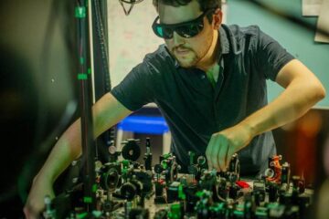
Researchers at Martinos Center for Biomedical Imaging at Massachusetts General Hospital in Boston helped develop a combined optical/x-ray imaging system capable of obtaining both structural and functional information of the breast.
The two technologies used were digital breast tomosynthesis (DBT), a three-dimensional application of digital mammography, and diffuse optical tomography (DOT), which measures levels of hemoglobin concentration, oxygen saturation and other cellular characteristics, based on how light from a near-infrared laser is absorbed and scattered within tissue.
“By co-registering optical and x-ray data, radiologists are able to map suspicious findings and analyze the functional characteristics of those areas,” said lead researcher Qianqian Fang, Ph.D., a radiology instructor at Harvard Medical School.
In the study, combined DBT and DOT was performed on 189 breasts from 125 women with an average age of 56 years. To perform the procedure, an optical source and detector probes were attached to a DBT unit and, with the breast compressed, optical data was acquired. The optical probes were then removed without altering the breast compression and a DBT scan was performed.
“We are very excited about adding optical imaging to DBT, because it is low-cost, safe, noninvasive and fast,” Dr. Fang said.
Of the 189 imaging studies, 138 were negative, and 51 showed evidence of lesions. As determined by breast biopsy, 26 lesions of the 51 lesions were malignant, and 25 were benign.
In the 26 malignant tumors, total hemoglobin concentration (HbT) was significantly greater than in the normal glandular tissue of the same breast. Solid benign lesions and cysts had significantly lower HbT contrast compared to the malignant lesions.
“By providing additional differentiation of malignant and benign lesions, combined optical and x-ray imaging could potentially reduce unnecessary biopsies,” Dr. Fang said.
In the study, oxygen saturation levels were significantly lower in cysts compared to those in malignant and solid benign lesions and glandular breast tissue.
“Although cysts are easy to diagnose using ultrasound, distinguishing cysts from malignant or benign lesions during a mammogram would save women the anxiety and costs associated with a second procedure,” Dr. Fang said. “We are hopeful that this combined system may help improve the efficiency and diagnostic accuracy of breast screening.”
The study is part of an ongoing research effort to improve breast cancer diagnosis led by Daniel B. Kopans, M.D., and David Boas, Ph.D., and funded by the National Institutes of Health.
“Combined Optical and X-ray Tomosynthesis Breast Imaging.” Collaborating with Dr. Fang were Juliette Selb, Ph.D., Stefan A. Carp, Ph.D., Greg Boverman, Ph.D., Eric L. Miller, Ph.D., Dana H. Brooks, Ph.D., Richard H. Moore, B.S., Daniel B. Kopans, M.D., and David A. Boas, Ph.D.
Radiology is edited by Herbert Y. Kressel, M.D., Harvard Medical School, Boston, Mass., and owned and published by the Radiological Society of North America, Inc. (http://radiology.rsna.org/)
RSNA is an association of more than 46,000 radiologists, radiation oncologists, medical physicists and related scientists committed to excellence in patient care through education and research. The Society is based in Oak Brook, Ill. (RSNA.org)
For patient-friendly information on breast imaging, visit RadiologyInfo.org.
Media Contact
More Information:
http://www.rsna.orgAll latest news from the category: Medical Engineering
The development of medical equipment, products and technical procedures is characterized by high research and development costs in a variety of fields related to the study of human medicine.
innovations-report provides informative and stimulating reports and articles on topics ranging from imaging processes, cell and tissue techniques, optical techniques, implants, orthopedic aids, clinical and medical office equipment, dialysis systems and x-ray/radiation monitoring devices to endoscopy, ultrasound, surgical techniques, and dental materials.


Newest articles
Combatting disruptive ‘noise’ in quantum communication
In a significant milestone for quantum communication technology, an experiment has demonstrated how networks can be leveraged to combat disruptive ‘noise’ in quantum communications. The international effort led by researchers…
Stretchable quantum dot display
Intrinsically stretchable quantum dot-based light-emitting diodes achieved record-breaking performance. A team of South Korean scientists led by Professor KIM Dae-Hyeong of the Center for Nanoparticle Research within the Institute for…

Internet can achieve quantum speed with light saved as sound
Researchers at the University of Copenhagen’s Niels Bohr Institute have developed a new way to create quantum memory: A small drum can store data sent with light in its sonic…

