Whole body MRI is highly accurate in the early detection of breast cancer metastases
Whole body MRI is a noninvasive medical test that helps physicians diagnose and treat breast cancer. Breast cancer cells commonly spread to the bones, lungs, liver, or brain. Metastatic breast cancer tumors may be found before or at the same time as the primary tumor, or months and even years later.
“It is important that we detect metastases early in order to ensure the patient is getting the appropriate treatment. This study shows that whole body MRI can accomplish this task and is ready to be used for this indication,” said Joshita Singh, MD, lead author of the study.
Besides MRI, other imaging modalities commonly used to detect breast cancer metastases include positron emission tomography – computed tomography (PET/CT), chest X-rays, bone scans, and ultrasound of the abdomen and pelvis.
The study, performed at Deenanath Mangeshkar Hospital and Research Center in Pune, India, included 99 patients with known breast cancer who were evaluated for metastases using whole body MRI. “Of the 99 patients, MRI accurately revealed that 47 patients were positive for metastases while 52 were negative. Of those patients who were positive for metastases, whole body MRI frequently detected bone metastases earlier when the patient was still asymptomatic,” said Singh.
“As our study suggests, whole body MRI is an effective tool for the detection of metastases and unlike other procedures commonly used in this role, it emits no radiation. We highly recommend that whole body MRI be the imaging modality of choice for the detection of metastases in patients with breast cancer,” she said.
This study will be presented on Thursday, May 6 at 11:20 a.m. Pacific Time. For a copy of the abstract or to schedule an interview with Dr. Singh, please contact Heather Curry via E-MAIL at hcurry@acr-arrs.org.
About ARRS
The American Roentgen Ray Society (ARRS) was founded in 1900 and is the oldest radiology society in the United States. Its monthly journal, the American Journal of Roentgenology, began publication in 1906. Radiologists from all over the world attend the ARRS annual meeting to participate in instructional courses, scientific paper presentations and scientific and commercial exhibits related to the field of radiology. The Society is named after the first Nobel Laureate in Physics, Wilhelm Röentgen, who discovered the x-ray in 1895.
Media Contact
More Information:
http://www.arrs.orgAll latest news from the category: Medical Engineering
The development of medical equipment, products and technical procedures is characterized by high research and development costs in a variety of fields related to the study of human medicine.
innovations-report provides informative and stimulating reports and articles on topics ranging from imaging processes, cell and tissue techniques, optical techniques, implants, orthopedic aids, clinical and medical office equipment, dialysis systems and x-ray/radiation monitoring devices to endoscopy, ultrasound, surgical techniques, and dental materials.


Newest articles

Wildfire danger to increase due to climate change
WSL Institute for Snow and Avalanche Research (SLF) researchers expect an elevated wildfire danger in the Alpine Foreland from 2040 onwards due to changing meteorological conditions. The danger currently remains…
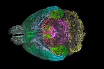
Advanced Brain Science Without Coding Expertise
Researchers at Helmholtz Munich and the LMU University Hospital Munich introduce DELiVR, offering a new AI-based approach to the complex task of brain cell mapping. The deep learning tool democratizes…
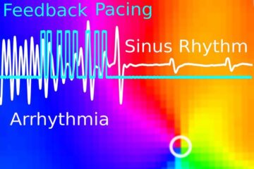
Gentle defibrillation for the heart
Using light pulses as a model for electrical defibrillation, Göttingen scientists developed a method to assess and modulate the heart function. The research team from the Max Planck Institute for…

