Bern Software Analyses Brain Tumours at Lightning Speed
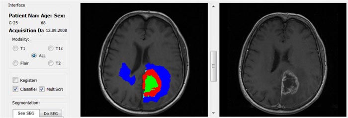
A screenshot of BraTumIA in action. (Mauricio Reyes)
The fully automatic computer program requires a maximum of 10 minutes per patient for the analysis of the magnetic resonance image of a brain tumour. A doctor requires between 30 and 60 minutes, however.
As the first software worldwide to do so, BRaTumIA also examines the tumour on a three dimensional basis without the need for human support, a task a doctor would take a far longer time to complete, and with a higher risk of error. As a result, manual daily hospital measurements are now only
occurring on two levels.
The program was developed and clinically tested by a team of doctors and engineers from the Bern University Hospital and the Institute for Surgical Technology and Biomechanics (ISTB) at the University of Bern, under the leadership of professors Roland Wiest (University Institute for Neuroradiology) and Mauricio Reyes (ISTB).
Analysis Down to the Tiniest Detail
BraTumIA provides Neuroradiologists with the optimum support in their analysis. The software compares the patient's MRI images with all of the previously gathered statistical data and determines the tissue structures of the malignant tumour right down to the tiniest detail. Prof. Roland Wiest, Neuroradiologist and Leader of the Support Centre of Advanced Neuroimaging at the Bern University Hospital:
“The precision segmenting of the tumour tissue is enabling us to use the image information to optimise the treatment on an ever more precise basis. This is hugely important, as new treatment strategies for gliomas – malignant tumours – can receive exact information on the growth data
concerning the tumours.”
Data Mining: Learning Software
It is in this context that BraTumIA has joined the medical data mining trend. Similar to conventional data mining, in which data regarding customers' buying and reading habits are gathered in the internet, the software consistently improves through the constant gathering of new statistical data. BraTumIA is currently the focus of considerable international attention: the Washington Post used the software at the beginning of October to demonstrate data mining in the analytical medical sector – in international comparisons, the software has consistently achieved a top three position in terms of its measuring accuracy.
The possibility of being able to continuously 'feed' the software with new statistical data is decisive for future brain tumour patients: when doctors analyse MRI images manually, analytical errors are theoretically possible in several different directions. The software, however, only makes analytical errors in the same direction, if at all. Doctors can check any such errors on a quick and targeted basis and reduce them to the minimum.
Soon to be Available for MS and Strokes
The intensive collaboration of the engineers and doctors in the development of BraTumIA could soon be benefitting to patients suffering from multiple sclerosis (MS) and those who have suffered stroke. The research group is currently working at full speed on two further versions of the software.
With MS patients, BraTumIA is set to provide precise analyses of inflamed brain tissue in the white brain matter (plaques). With stroke patients the software will serve the purpose of risk analysis: In the immediate aftermath of a stroke it recognises which parts of the brain are likely to remain damaged subsequent to treatment. In this task, BraTumIA also makes use of clinically gathered data.
The research on the computer supported analysis using BraTumIA at the Bern University Hospital and the University of Bern is being supported by the European Union, the Swiss National Fund and the Bern and Swiss Cancer League.
Weitere Informationen:
http://www.plosone.org/article/info%3Adoi%2F10.1371%2Fjournal.pone.0096873
https://hal.inria.fr/hal-00935640/PDF/brats_preprint.pdf
http://www.washingtonpost.com/blogs/innovations/wp/2014/10/01/the-incredible-potential-and-dangers-of-data-mining-health-records/
Media Contact
All latest news from the category: Medical Engineering
The development of medical equipment, products and technical procedures is characterized by high research and development costs in a variety of fields related to the study of human medicine.
innovations-report provides informative and stimulating reports and articles on topics ranging from imaging processes, cell and tissue techniques, optical techniques, implants, orthopedic aids, clinical and medical office equipment, dialysis systems and x-ray/radiation monitoring devices to endoscopy, ultrasound, surgical techniques, and dental materials.


Newest articles

Superradiant atoms could push the boundaries of how precisely time can be measured
Superradiant atoms can help us measure time more precisely than ever. In a new study, researchers from the University of Copenhagen present a new method for measuring the time interval,…

Ion thermoelectric conversion devices for near room temperature
The electrode sheet of the thermoelectric device consists of ionic hydrogel, which is sandwiched between the electrodes to form, and the Prussian blue on the electrode undergoes a redox reaction…

Zap Energy achieves 37-million-degree temperatures in a compact device
New publication reports record electron temperatures for a small-scale, sheared-flow-stabilized Z-pinch fusion device. In the nine decades since humans first produced fusion reactions, only a few fusion technologies have demonstrated…

