“Windows” into the Cell’s Interior – New Method Enables Deeper Insights into the Cell
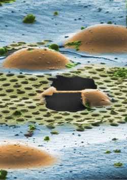
'Shock frozen' cell after treatment with the ion beam. Graphic: Alexander Rigort & Felix Bäuerlein / Copyright: MPI of Biochemistry<br>
However, with this method only very small cells or thin peripheral regions of larger cells can be investigated directly. Scientists of the Max Planck Institute of Biochemistry (MPIB) in Martinsried near Munich have now developed a procedure to provide access to cellular regions which were previously nearly inaccessible.
Using focused ion beam (FIB) technology, specific cellular material can be cut out, opening up thin “windows” into the cell’s interior. This alternative approach enables the preparation of larger cellular samples devoid of artefacts. The study was recently published in PNAS USA.
With cryo-electron tomography, pioneered by the Department of Molecular Structural Biology headed by Wolfgang Baumeister, researchers can now directly analyze three-dimensional cellular structures. The entire cell or individual cell components are “shock frozen” and enclosed in glass-like ice, thus preserving their spatial structure. The transmission electron microscope then enables the acquisition of two-dimensional projections from different perspectives. Finally, the scientists reconstruct a high-resolution three-dimensional volume from these images. However, the electron beam can penetrate only very thin specimens (for example bacteria cells) up to a thickness of 500 nanometers. Cells of higher organisms are clearly thicker. State-of-the-art electron microscopic preparation techniques are therefore necessary to make also larger objects accessible for cryo-electron tomography.
“The artefact-free and, in particular, targeted preparation of larger cells is a critical step,” explained Alexander Rigort, MPIB scientist. “With the traditional methods, we could never rule out that structures we wanted to investigate were changed.” The meaningfulness of the results was therefore limited, according to the biologist.
Using a focused ion beam microscope (FIB), researchers can now mill single layers of the frozen-hydrated cell and remove them in a controlled manner – thus rendering thin, tailor-made electron-transparent “windows”. An additional advantage of ion thinning is that mechanical sectioning artefacts are completely avoided. This method was originally developed for the material sciences. In structural biology the method shall now provide deeper insights into the molecular organization of the cell’s interior. The thinner the “windows” are, the higher the attainable resolution in the electron microscope. “Now precise insights into the macromolecular architecture of cell regions are possible that were previously nearly inaccessible for cryo-electron microscopy,” said Jürgen Plitzko, scientist at the MPIB.
Original Publication
A. Rigort, F. Bäuerlein, E. Villa, M. Eibauer, T. Laugks, W. Baumeister and J. M. Plitzko: Focused Ion Beam micromachining of eukaryotic cells for cryoelectron tomography. Proc. Natl. Acad. Sci. USA, March 5, 2012
Doi:10.1073/pnas.1201333109.
Contact
Dr. Jürgen M. Plitzko
Molecular Structural Biology
Max Planck Institute of Biochemistry
Am Klopferspitz 18
82152 Martinsried
Germany
E-Mail: plitzko@biochem.mpg.de
http://www.biochem.mpg.de/baumeister
Dr. Alexander Rigort
Molecular Structural Biology
Max Planck Institute of Biochemistry
Am Klopferspitz 18
82152 Martinsried
Germany
E-Mail: rigort@biochem.mpg.de
Anja Konschak
Public Relations
Max Planck Institute of Biochemistry
Am Klopferspitz 18
82152 Martinsried
Germany
Phone: +49 (0) 89 8578-2824
E-Mail: konschak@biochem.mpg.de
Media Contact
All latest news from the category: Life Sciences and Chemistry
Articles and reports from the Life Sciences and chemistry area deal with applied and basic research into modern biology, chemistry and human medicine.
Valuable information can be found on a range of life sciences fields including bacteriology, biochemistry, bionics, bioinformatics, biophysics, biotechnology, genetics, geobotany, human biology, marine biology, microbiology, molecular biology, cellular biology, zoology, bioinorganic chemistry, microchemistry and environmental chemistry.


Newest articles
Combatting disruptive ‘noise’ in quantum communication
In a significant milestone for quantum communication technology, an experiment has demonstrated how networks can be leveraged to combat disruptive ‘noise’ in quantum communications. The international effort led by researchers…
Stretchable quantum dot display
Intrinsically stretchable quantum dot-based light-emitting diodes achieved record-breaking performance. A team of South Korean scientists led by Professor KIM Dae-Hyeong of the Center for Nanoparticle Research within the Institute for…
Internet can achieve quantum speed with light saved as sound
Researchers at the University of Copenhagen’s Niels Bohr Institute have developed a new way to create quantum memory: A small drum can store data sent with light in its sonic…

