Trinity biochemists devise snappy new technique for blueprinting cell membrane proteins
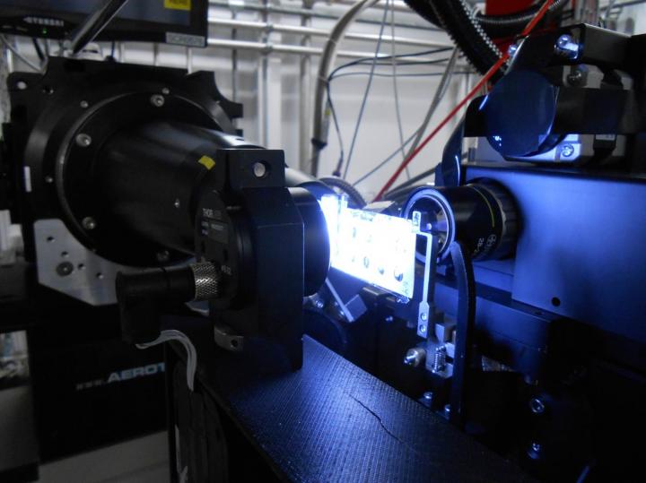
The in meso in situ serial crystallography (IMISX) method allows researchers to quickly and accurately blueprint the 3-D structure of proteins. Credit: Martin Caffrey (Trinity College Dublin).
The breakthrough will make a big splash in the field of drug discovery and development, where precise protein structure blueprints can help researchers understand how individual proteins work. Critically, these blueprints can show weaknesses that allow drug developers to draw up specific battle plans in the fight against diseases and infections.
Professor of Membrane Structural and Functional Biology at Trinity, Martin Caffrey, is the senior author of the research, which has just been published in the international peer-reviewed journal Acta Crystallographica D. He said:
“This is a truly exciting development. We have demonstrated the method on a variety of cell membrane proteins, some of which act as transporters. It will work with existing equipment at a host of facilities worldwide, and it is very simple to implement.”
Over 50% of drugs on the market target cell membrane proteins, which are vital for the everyday functioning of complex cellular processes. They act as transporters to ensure that specific molecules enter and leave our cells, as signal interpreters important in decoding messages and initiating responses, and as agents that speed up appropriate responses.
The major challenge facing researchers is the production of large membrane protein crystals, which are used to determine the precise 3-D structural blueprints. That challenge has now been lessened thanks to the Trinity biochemists' advent – the in meso in situ serial crystallography (IMISX) method.
Beforehand, researchers needed to harvest protein crystals and cool them at inhospitable temperatures in a complex set of events that was damaging, inefficient and prone to error. The IMISX method allows researchers to determine structural blueprints as and where the crystals grow.
Professor Caffrey added: “The best part of this is that these proteins are as close to being 'live' and yet packaged in the crystals we need to determine their structure as they could ever be. As a result, this breakthrough is likely to supplant existing protocols and will make the early stages of drug development considerably more efficient.”
The work was done in collaboration with scientists at the Swiss Light Source and the University of Konstanz and was supported by a grant from Science Foundation Ireland.
###
A pdf is available on request.
Media Contact
All latest news from the category: Life Sciences and Chemistry
Articles and reports from the Life Sciences and chemistry area deal with applied and basic research into modern biology, chemistry and human medicine.
Valuable information can be found on a range of life sciences fields including bacteriology, biochemistry, bionics, bioinformatics, biophysics, biotechnology, genetics, geobotany, human biology, marine biology, microbiology, molecular biology, cellular biology, zoology, bioinorganic chemistry, microchemistry and environmental chemistry.


Newest articles

High-energy-density aqueous battery based on halogen multi-electron transfer
Traditional non-aqueous lithium-ion batteries have a high energy density, but their safety is compromised due to the flammable organic electrolytes they utilize. Aqueous batteries use water as the solvent for…

First-ever combined heart pump and pig kidney transplant
…gives new hope to patient with terminal illness. Surgeons at NYU Langone Health performed the first-ever combined mechanical heart pump and gene-edited pig kidney transplant surgery in a 54-year-old woman…

Biophysics: Testing how well biomarkers work
LMU researchers have developed a method to determine how reliably target proteins can be labeled using super-resolution fluorescence microscopy. Modern microscopy techniques make it possible to examine the inner workings…

