The large-scale stability of chromosomes
Cromatin. Credit: SISSA
“Interphase” refers to the period in the cell cycle in which chromosomes spend most of their time. During this phase, in between mitoses, chromosomes live “dissolved'' in the nucleus where they carry out the processes required for the duplication of genetic material.
Our current knowledge regarding the behaviour of chromosomes during interphase is unfortunately quite limited; for example, we would need to know more about the three-dimensional structure of the chromatin filament – the long molecule that makes up the chromosomes and that consists of DNA and other proteins – and how it changes in time and space. The shape of the chromosome is in fact important for its function as it allows or prevents access to portions of genetic code for the duplication processes.
In addition to experimental observation, another important method of study is computer simulation, based on theoretical models of chromatin. A new study has extended work done previously, which used a simpler model of chromatin consisting up of a single fibre.
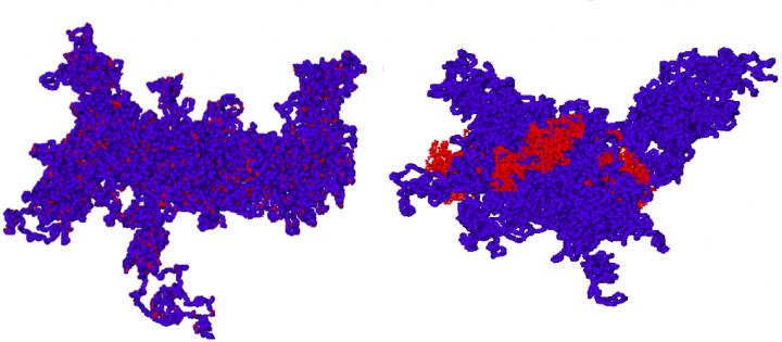
In the new study, the filament could be made up of two types of fibre, one thicker and one thinner, in varying proportions. Experimental studies have indeed demonstrated the existence of two main types of chromatin, with thicknesses of 10 or 30 nm.
In the study, the scientists made molecular dynamics simulations for model chromosomes in various conditions: they added increasingly large amounts of 10 nm fibre to the homopolymer chromatin, consisting of the stiffer 30 nm fibre only.
The model used in the new study, while a simplification of the real molecule, makes the simulation more realistic. The aim of the study was to evaluate whether small-scale modifications in the chromatin fibre lead to large-scale changes in the behavior of the chromosome.
“Even after the introduction of the more flexible second fibre, the chromosome remains spatially stable” explains Ana-Maria Florescu, first author of the study and SISSA research scientist. “More in detail”, adds Angelo Rosa, SISSA research fellow who coordinated the work, “we found that reorganization occurs only on spatial scales below 0.1 Mbp (million base pairs) and on time scales shorter than a few seconds”.
One significant implication of this study concerns the techniques used for the experimental observation of the fibres: those most commonly used today have inadequate resolution to be able to observe this type of reorganization, so we aim to develop new methodologies (like the FISH technique based on oligonucleotides) able to visualize even genome distances smaller than 0.1 Mbp.
Media Contact
All latest news from the category: Life Sciences and Chemistry
Articles and reports from the Life Sciences and chemistry area deal with applied and basic research into modern biology, chemistry and human medicine.
Valuable information can be found on a range of life sciences fields including bacteriology, biochemistry, bionics, bioinformatics, biophysics, biotechnology, genetics, geobotany, human biology, marine biology, microbiology, molecular biology, cellular biology, zoology, bioinorganic chemistry, microchemistry and environmental chemistry.


Newest articles

High-energy-density aqueous battery based on halogen multi-electron transfer
Traditional non-aqueous lithium-ion batteries have a high energy density, but their safety is compromised due to the flammable organic electrolytes they utilize. Aqueous batteries use water as the solvent for…

First-ever combined heart pump and pig kidney transplant
…gives new hope to patient with terminal illness. Surgeons at NYU Langone Health performed the first-ever combined mechanical heart pump and gene-edited pig kidney transplant surgery in a 54-year-old woman…

Biophysics: Testing how well biomarkers work
LMU researchers have developed a method to determine how reliably target proteins can be labeled using super-resolution fluorescence microscopy. Modern microscopy techniques make it possible to examine the inner workings…

