The amazing flexibility of red blood cells
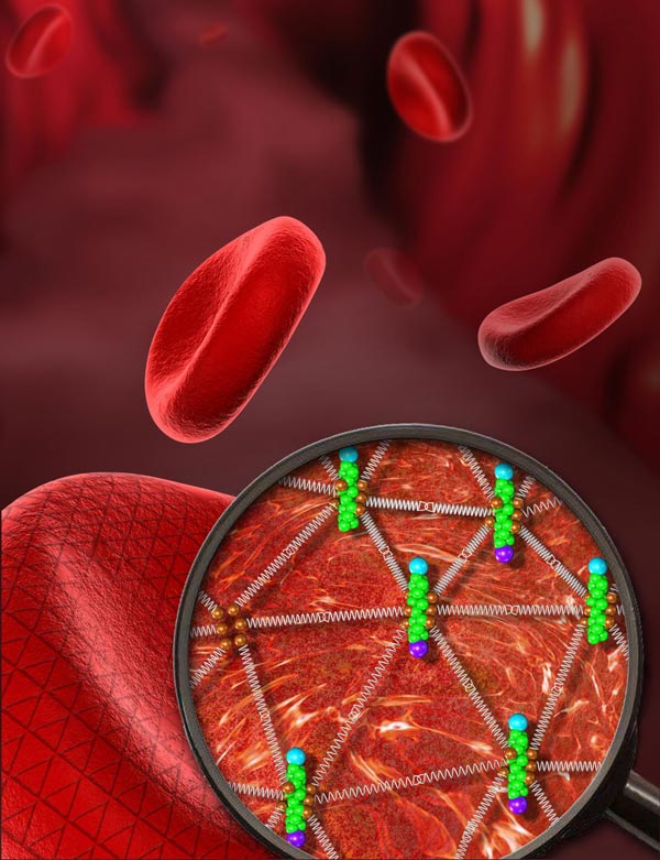
A super-resolution microscopy technique called STORM reveals the two-dimensional triangular protein meshwork that underlies the membrane of the red blood cell and is key to its flexibility. Credit: Ke Xu, UC Berkeley
One of today's sharpest imaging tools, super-resolution microscopy, produces sparkling images of what until now has been the blurry interior of cells, detailing not only the cell's internal organs and skeleton, but also providing insights into cells' amazing flexibility.
In the current issue of the journal Cell Reports, Ke Xu and his colleagues at UC Berkeley use the technique to provide a sharp view of the geodesic mesh that supports the outer membrane of a red blood cell, revealing why such cells are sturdy yet flexible enough to squeeze through narrow capillaries as they carry oxygen to our tissues.
The discovery could eventually help uncover how the malaria parasite hijacks this mesh, called the sub-membrane cytoskeleton, when it invades and eventually destroys red blood cells.
“People know that the parasite interacts with the cytoskeleton, but how it does it is unclear because there has been no good way to look at the structure,” said Xu, an assistant professor of chemistry. “Now that we have resolved what is really going on in a normal healthy cell, we can ask what changes under infection with parasites and how drugs affect the interaction.”
Typical human cells have a two-dimensional skeleton that supports the outer membrane and a three-dimensional interior skeleton that supports all the organelles inside and serves as a transportation system throughout the cell.
Red blood cells, however, have only the membrane supports and no internal scaffolding, so they're basically a balloon filled with molecules of oxygen-carrying hemoglobin. Because of their simpler structure, red blood cells are ideal for studying the skeleton that supports the membrane in all cells.
Electron microscope images earlier showed that the sub-membrane cytoskeleton in red blood cells is a triangular mesh of proteins, reminiscent of a geodesic dome. But measurements of the size of the triangular subunits were made by flattening out the domed membrane of a dead and dried-out cell, which distorts the structure.
STORMing the cytoskeleton
Xu was a postdoctoral fellow in the Harvard University lab of one of the inventors of super-resolution microscopy, Xiaowei Zhuang, and is an expert on the version called STORM (stochastic optical reconstruction microscopy). Super-resolution microscopy gives about 10 times better resolution than standard light microscopy and works well with wet and live cells.
Using STORM, Xu, former Berkeley postdoc Leiting Pan and graduate student Rui Yan were able to image the full sub-membrane cytoskeleton of fresh red blood cells and discovered that the triangles of the mesh are about half the size of found in earlier measurements done with electron microscopy: each side is 80 nanometers long, instead of 190 nanometers.
The distinction is critical: The building blocks of the mesh are a protein called spectrin, which can be stretched to a maximum of about 190 nanometers in length. If the mesh were made of stretched spectrin, it would be rigid, Xu said. But since its normal length is a relaxed 80 nanometers, it acts like a spring. “It is more like a spring in its relaxed state, where it has much flexibility under compression or stretching, so that gives red blood cells a lot of elasticity under different physiological conditions, such as squeezing through a narrow capillary,” Yan said.
At the vertices of the mesh, where five to six spectrin proteins come together, is a different protein: actin. Actin is a standard part of the sub-membrane cytoskeleton and one of the main structural components of the cell.
Tears in the mesh
Interestingly, STORM revealed never-before-seen holes in the cytoskeletal mesh that may also be critical to its flexibility.
“This is a defect in the network, but there might be a reason for it,” said Xu, who is also a Chan Zuckerberg Biohub Investigator. “The cell would want to change structure rapidly as it goes through the capillaries, and having those defects is helpful in reorganizing the shape without breaking the mesh. It can act as a weak point as they try to squeeze through things, they can start to bend around those points.”
Xu actually discovered the key structural role of spectrin. While still at Harvard, he used STORM to look at the skeletal structure of neurons, and discovered that actin proteins form precisely spaced rings along the entire length of the axon – which can be as much as a foot long – much like the ribs of a snake. They are separated by exactly 190 nanometers, and when he looked through textbooks for proteins with that length, he came across spectrin. He subsequently used STORM to confirm that in its stretched state, spectrin proteins are the spacers between the rings, keeping them precisely separated.
“The ringed skeleton makes the axon a very stable but bendable structure,” Xu said, whereas the regular spacing may be key to its electrical conductivity.
Super-resolution microscopy employs a trick to overcome the diffraction limit of light microscopy, which prevents conventional light microscopes from resolving things smaller than half the size of the wavelength of the light, which for visible light is about 300 nanometers.
STORM involves attaching a blinking light source to individual molecules and then isolating each light's position independently of the others, building up a complete image much like the 1880s artists who developed pointillism, producing images from individual dots of paint.
Typically chemists attach these flashing sources to all molecules of the same type in a cell, such as all actin molecules, but since only a small percentage of the sources blink on at any one time, it's possible to pinpoint the exact location of each. Today's best resolution is about 10 nanometers, Xu said, which is about the size of a single protein or molecule.
###
The work was supported by the National Natural Science Foundation of China, a Pew Biomedical Scholars Award and a Packard Fellowship for Science and Engineering. Coauthor and postdoc Wan Li contributed to experimental design and data analysis.
Media Contact
All latest news from the category: Life Sciences and Chemistry
Articles and reports from the Life Sciences and chemistry area deal with applied and basic research into modern biology, chemistry and human medicine.
Valuable information can be found on a range of life sciences fields including bacteriology, biochemistry, bionics, bioinformatics, biophysics, biotechnology, genetics, geobotany, human biology, marine biology, microbiology, molecular biology, cellular biology, zoology, bioinorganic chemistry, microchemistry and environmental chemistry.


Newest articles

Floating solar’s potential
… to support sustainable development by addressing climate, water, and energy goals holistically. A new study published this week in Nature Energy raises the potential for floating solar photovoltaics (FPV)…

Skyrmions move at record speeds
… a step towards the computing of the future. An international research team led by scientists from the CNRS1 has discovered that the magnetic nanobubbles2 known as skyrmions can be…
A flexible and efficient DC power converter for sustainable-energy microgrids
A new DC-DC power converter is superior to previous designs and paves the way for more efficient, reliable and sustainable energy storage and conversion solutions. The Kobe University development can…

