Sweetening neurotransmitter receptors and other neuronal proteins
Many neuronal proteins have atypical glycosylation profiles consistent with the virtual absence of an important organelle, the Golgi apparatus, in neuronal processes Max Planck Institute for Brain Research
To rapidly carry information throughout the body, neurons form intricate networks by sending long protrusions to physically contact other neurons, sometimes meters away from where their main body (hence called the cell body) is located. These tree-like protrusions are either called axons if they are used to send information or dendrites if they receive information from other neurons.
Axons and dendrites contact one another at specialized communication structures called synapses where the axon stimulates the dendrite by releasing small chemical compounds called neurotransmitters, that bind to specialized proteins called neurotransmitter receptors which are expressed at the surface of dendrites, and trigger electric signals that travel through the rest of the cell when they are activated.
Sugar molecules, so-called glycans, are one of the components of these receptor proteins, but were not well studied in neurons until now. In a recent paper, Cyril Hanus and colleagues in the Schuman Lab at the Frankfurt Max Planck Institute for Brain Research showed that many neuronal proteins have atypical glycosylation profiles consistent with the virtual absence of an important organelle, the Golgi apparatus, in neuronal processes. These atypical sugar molecules change how the receptors respond to neurotransmitters.
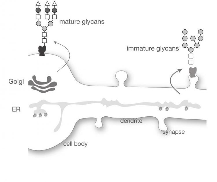
In all cells, surface proteins are made inside the cell in a compartment called the endoplasmic reticulum. During this process, one or several complex sugar molecules are usually added to newly made proteins. These sugar molecules are then modified as the proteins leave the endoplasmic reticulum and pass through another compartment called the Golgi apparatus on the way to the cell surface. The precise number and structure of the sugar molecules attached to the protein define its glycosylation profile.
To adapt their response to synaptic stimulation, for example during learning, neurons control how many receptors are expressed at the cell surface, in particular by producing more of these receptors. These new receptors can be made in the main cell body but, to speed up the process during memory formation, also locally in dendrites, close to synapses where they are needed. However, while the endoplasmic reticulum is found all over the neuron, including in the dendrites, the Golgi apparatus is generally only present in the main cell body. It was not known how surface proteins are made in the dendrites or how the proteins’ glycosylation profiles are altered in the absence of a Golgi apparatus.
Hanus et al. used microscopy and advanced biochemical techniques to study the glycosylation profiles of surface proteins in rat neurons. The experiments revealed that immature glycosylation profiles are found on hundreds of different proteins that have been transported to the cell surface. This includes many neurotransmitter receptors but also numerous other key surface proteins. Next, Hanus et al. selectively blocked the modification of sugar molecules on proteins in the Golgi apparatus. This showed that dendrites are able to form and work properly even if surface proteins have primarily immature glycosylation profiles. Finally, the authors showed that these immature glycosylation profiles change the way neurotransmitter receptors react to stimulation by neurotransmitters, showing that the glycosylation profile of surface proteins impact their function.
Cyril Hanus: “These new results show that neurons can produce surface proteins in a way that is different from other cells, and in doing so control important aspects of the function of these proteins, a process that may, in the long run, be exploited to design new medicines tailored to neuronal proteins.”
Publication: Hanus, C., Geptin, H., Tushev, G., Garg, S., Alvarez-Castelao, B., Sambandan, S., Kochen, L., Hafner, A.S., Langer, J.D., Schuman, E.M. (2016). Unconventional secretory processing diversifies neuronal ion channel properties. eLife 5: e20609 (https://elifesciences.org/content/5/e20609/article-info)
Media Contact
All latest news from the category: Life Sciences and Chemistry
Articles and reports from the Life Sciences and chemistry area deal with applied and basic research into modern biology, chemistry and human medicine.
Valuable information can be found on a range of life sciences fields including bacteriology, biochemistry, bionics, bioinformatics, biophysics, biotechnology, genetics, geobotany, human biology, marine biology, microbiology, molecular biology, cellular biology, zoology, bioinorganic chemistry, microchemistry and environmental chemistry.


Newest articles

Properties of new materials for microchips
… can now be measured well. Reseachers of Delft University of Technology demonstrated measuring performance properties of ultrathin silicon membranes. Making ever smaller and more powerful chips requires new ultrathin…

Floating solar’s potential
… to support sustainable development by addressing climate, water, and energy goals holistically. A new study published this week in Nature Energy raises the potential for floating solar photovoltaics (FPV)…

Skyrmions move at record speeds
… a step towards the computing of the future. An international research team led by scientists from the CNRS1 has discovered that the magnetic nanobubbles2 known as skyrmions can be…

