Staying in Shape
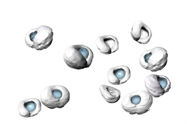
Manually segmented zebrafish retinas (grey) and lenses (blue) at different developmental stages. Tissue scaling during retinal growth is enabled by timely tissue-wide cell elongation. Matejčić / Norden, MPI-CBG
During the early development of an embryo, many tissues and organs already form their final shape. This shape has to be maintained while the organism keeps growing. As the correct shape of a tissue is often essential for its proper function, it is crucial to understand how it remains unchanged while the organism grows and develops.
So far, the interaction between cells inside tissues that enable growth while keeping shape are still poorly understood. Scientists at the Max Planck Institute of Molecular Cell Biology and Genetics (MPI-CBG) in Dresden in collaboration with Guillaume Salbreux from the Francis Crick Institute, present a 3D study of eye growth over development.
They were able to show that elongated cells are the key to maintain the shape of the retinal tissue during growth of a zebrafish. This concept could also apply to other organisms. The researchers published their findings in the journal PLOS Biology.
Understanding how tissues properly form and grow during the development of an organism is an important question in biology. Many tissues establish their shape early in development and thus need to maintain this shape as the tissue grows, similar to a balloon, which keeps the same form when you blow it up. This is the case for many human tissues, such as the nose or the eye.
The correct shape of a tissue or organ is often essential for its function, so it is crucial to understand for example how a tiny nose can maintain its shape while it is growing. The behaviors of cells underlying such coordinated growth still need to be identified. Previous studies mostly explored tissue growth and shape in two dimensions. Thus, an evaluation of tissue growth in three dimensions is still needed to fully comprehend shape and size.
About the MPI-CBG
The Max Planck Institute of Molecular Cell Biology and Genetics (MPI-CBG) is one of 84 institutes of the Max Planck Society, an independent, non-profit organization in Germany. 500 curiosity-driven scientists from over 50 countries ask: How do cells form tissues? The basic research programs of the MPI-CBG span multiple scales of magnitude, from molecular assemblies to organelles, cells, tissues, organs, and organisms.
The team around MPI-CBG research group leader Dr. Caren Norden in collaboration with Guillaume Salbreux, a former colleague from the Max Planck Institute for the Physics of Complex Systems and now at the Francis Crick Institute, set out to explore these important, exciting questions and took advantage of the exceptional imaging possibilities that exist for the developing zebrafish. A tissue that was particularly suitable for this study is the retinal neuroepithelium, an important part of the central nervous system which forms a cup with a smooth surface early in development.
Later, while still keeping its shape, this neuroepithelium forms a neuronal tissue, that will transmit the light impulses from the eye to the brain. Thus, for the retina, it is particularly important to form with an undisturbed shape, so light can pass through it smoothly. The first author of the study, Dr. Marija Matejčić explains: “We showed that cells that build the retina need to elongate to maintain the shape of the tissue unchanged while it grows. Retinal cells elongate together, after redistributing an inner component at the same time throughout the tissue. In this way, the cells and the tissue stay in great shape!”
The protein actin plays a crucial role in this process: a redistribution of actin at a right time makes sure that the height of cells can increase. If, on the other hand, the actin redistribution is blocked, the cell height does not increase, which leads to a disturbed, folded tissue shape of the otherwise smooth retinal tissue which would interfere with organ function
“This study provides an important example of how growth of a tissue that needs to maintain its form is achieved through changes of cell shape,” says Dr. Caren Norden, who oversaw the study. The concept could also apply to other organisms or the increasingly popular organoids. Dr. Norden adds: “The next challenge will be to expand investigations to growth phenomena in organoid systems, miniature, simple and lab-grown versions of an organ, including human organoids. This will deepen our understanding of developmental programs in model organisms and humans.”
Caren Norden
+49 (0) 351 210 2802
norden@mpi-cbg.de
Marija Matejčić, Guillaume Salbreux, Caren Norden
A non-cell-autonomous actin redistribution enables isotropic retinal growth
PLoS Biol, August 10, 2018. https://doi.org/10.1371/journal.pbio.2006018
Media Contact
More Information:
https://www.mpi-cbg.de/de/home/All latest news from the category: Life Sciences and Chemistry
Articles and reports from the Life Sciences and chemistry area deal with applied and basic research into modern biology, chemistry and human medicine.
Valuable information can be found on a range of life sciences fields including bacteriology, biochemistry, bionics, bioinformatics, biophysics, biotechnology, genetics, geobotany, human biology, marine biology, microbiology, molecular biology, cellular biology, zoology, bioinorganic chemistry, microchemistry and environmental chemistry.


Newest articles

Superradiant atoms could push the boundaries of how precisely time can be measured
Superradiant atoms can help us measure time more precisely than ever. In a new study, researchers from the University of Copenhagen present a new method for measuring the time interval,…

Ion thermoelectric conversion devices for near room temperature
The electrode sheet of the thermoelectric device consists of ionic hydrogel, which is sandwiched between the electrodes to form, and the Prussian blue on the electrode undergoes a redox reaction…

Zap Energy achieves 37-million-degree temperatures in a compact device
New publication reports record electron temperatures for a small-scale, sheared-flow-stabilized Z-pinch fusion device. In the nine decades since humans first produced fusion reactions, only a few fusion technologies have demonstrated…

