Fluorescent nanoparticles serve as flashlights in living cells

The ‘quantum dot’ nanoparticles used by Van Manen and Otto replace existing fluorescent labels that are employed to enable the cell’s biomolecules to light up under the microscope. While fluorescence microscopy continues to be instrumental in unraveling the intricate biological processes that take place inside living cells, it would be even more informative to combine it with the intracellular chemical analysis capabilities of vibrational spectroscopy techniques such as Raman microscopy.
Common fluorescent labels are not suitable for this combination, however, because the much stronger fluorescence overshadows the intrinsic weak Raman signals coming from cells. By taking fluorescent quantum dots that emit light in a wavelength region that is well-separated from Raman signals, the Dutch researchers now show that fluorescence microscopy can indeed be combined with Raman microscopy on the same cell.
Vibrations inside cells
Techniques based on vibrational spectroscopy are able to detect the specific vibrations that occur inside the cell’s biomolecules (such as DNA, proteins, and lipids), making them very powerful tools for ‘chemical fingerprinting’ of cells. In contrast to fluorescence microscopy, vibrational spectroscopy does not require the biomolecules of interest to be labeled, which is a great advantage. The Biophysical Engineering Group at the University of Twente, headed by prof. Vinod Subramaniam, has pioneered the application of Raman spectroscopy to investigate the chemical make-up of single cells, and this group is now worldwide at the front of high-resolution chemical mapping of cells by Raman microscopy.
In their Nano Letters article, the researchers demonstrate two applications of the hybrid fluorescence Raman technique. By illuminating white blood cells with UV light at a wavelength of 413 nm, the Raman signal from an enzyme that is critical in the innate immune response can be detected and visualized across the cell. The fluorescence signal of quantum dot nanoparticles that have been ingested by the cells can be visualized separately. The second application employs light at a wavelength of 647 nm, which results in the separate detection of Raman signals from cellular proteins and lipids and the fluorescence signal from the nanoparticles.
Van Manen and Otto expect that the fluorescence Raman microscopy combination will provide exciting new possibilities: the nanoparticles might be coated on their surface with antibodies against, for example, marker proteins for cancer cells. In this way the quantum dots will serve as a torch for specific cells, which can subsequently be subjected to a detailed chemical analysis by using Raman microscopy.
The research described in the Nano Letters article was funded by the Landsteiner Foundation for Blood Transfusion Research (Amsterdam, The Netherlands) and the MESA+ Institute for Nanotechnology at the University of Twente.
Media Contact
More Information:
http://bpe.tnw.utwente.nl/All latest news from the category: Life Sciences and Chemistry
Articles and reports from the Life Sciences and chemistry area deal with applied and basic research into modern biology, chemistry and human medicine.
Valuable information can be found on a range of life sciences fields including bacteriology, biochemistry, bionics, bioinformatics, biophysics, biotechnology, genetics, geobotany, human biology, marine biology, microbiology, molecular biology, cellular biology, zoology, bioinorganic chemistry, microchemistry and environmental chemistry.
Newest articles

Properties of new materials for microchips
… can now be measured well. Reseachers of Delft University of Technology demonstrated measuring performance properties of ultrathin silicon membranes. Making ever smaller and more powerful chips requires new ultrathin…

Floating solar’s potential
… to support sustainable development by addressing climate, water, and energy goals holistically. A new study published this week in Nature Energy raises the potential for floating solar photovoltaics (FPV)…

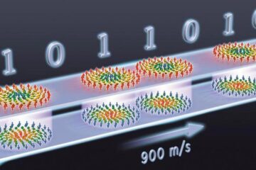
Skyrmions move at record speeds
… a step towards the computing of the future. An international research team led by scientists from the CNRS1 has discovered that the magnetic nanobubbles2 known as skyrmions can be…


