Molecular structure of the cell nucleoskeleton revealed for the first time
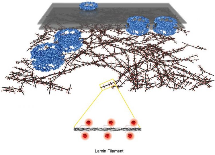
Nuclear lamina: architecture of delicate meshwork made of lamin filaments (filament rod in dark grey, globular domains in red) beneath nuclear membrane (transparent grey) and pore complexes (blue). Yagmur Turgay, University of Zurich
Compared to bacteria, in eukaryotes the genetic material is located in the cell nucleus. Its outer shell consists of the nuclear membrane with numerous nuclear pores. Molecules are transported into or out of the cell nucleus via these pores.
Beneath the membrane lies the nuclear lamina, a threadlike meshwork merely a few millionths of a millimeter thick. This stabilizes the cell nucleus and protects the DNA underneath from external influences. Moreover, the lamina plays a key role in essential processes in the cell nucleus – such as the organization of the chromosomes, gene activity and the duplication of genetic material before cell division.
Detailed 3D image of the nuclear lamina in its native environment
Now, for the first time, a team of researchers headed by cell biology professor Ohad Medalia from the Department of Biochemistry at UZH has succeeded in elucidating the molecular architecture of the nuclear lamina in mammalian cells in detail. The scientists studied fibroblast cells of mice using cryo-electron tomography.
“This technique combines electron microscopy and tomography, and enables cell structures to be displayed in 3D in a quasi-natural state,” explains Yagmur Turgay, the first author of the study. The cells are shock-frozen in liquid ethane at minus 190 degrees without being pretreated with harmful chemicals, thereby preserving the cell structures in their original state.
“The lamin meshwork is a layer that’s around 14 nanometers thick, located directly beneath the pore complexes of the nuclear membrane and consists of regions that are packed more or less densely,” says Yagmur Turgay, describing the architecture of the nucleoskeleton.
The scaffold is made of thin, threadlike structures that differ in length – the lamin filaments. Only 3.5 nanometers thick, the lamin filaments are much thinner and more delicate than the structures forming the cytoskeleton outside the cell nucleus in higher organisms.
New approach for research on progeria and muscular dystrophy
The building blocks of the filaments are two proteins – type A and B lamin proteins – which assemble into polymers. They consist of a long stem and a globular domain, much like a pin with a head. Individual mutations in the lamin gene elicit severe diseases with symptoms such as premature aging (progeria), muscle wasting (muscular dystrophy), lipodystrophy and damage of the nervous system (neuropathies).
“Cryo-electron tomography will enable us to study the structural differences in the nuclear lamina in healthy people and in patients with mutations in the lamin gene in detail in the future,” concludes Ohad Medalia. According to the structural biologist, this method permits the development of new disease models at molecular level, which paves the way for new therapeutic interventions.
Literature:
Yagmur Turgay, Matthias Eibauer, Anne E. Goldman, Takeshi Shimi, Maayan Khayat, Kfir Ben-Harush, Anna Dubrovsky-Gaupp, K. Tanuj Sapra, Robert D. Goldman, Ohad Medalia. The molecular architecture of lamins in somatic cells. Nature. March 1, 2017. DOI:10.1038/nature21382
Contact:
Prof. Dr. Ohad Medalia
Department of Biochemistry
University of Zurich
Phone: +41 44 635 55 22
E-mail: omedalia@bioc.uzh.ch
Dr. Yagmur Turgay
Department of Biochemistry
University of Zurich
Phone: +41 44 635 55 06
E-mail: y.turgay@bioc.uzh.ch
http://www.media.uzh.ch/en/Press-Releases/2017/structure-of-the-cell-nucleoskele…
Media Contact
All latest news from the category: Life Sciences and Chemistry
Articles and reports from the Life Sciences and chemistry area deal with applied and basic research into modern biology, chemistry and human medicine.
Valuable information can be found on a range of life sciences fields including bacteriology, biochemistry, bionics, bioinformatics, biophysics, biotechnology, genetics, geobotany, human biology, marine biology, microbiology, molecular biology, cellular biology, zoology, bioinorganic chemistry, microchemistry and environmental chemistry.


Newest articles

Biophysics: Testing how well biomarkers work
LMU researchers have developed a method to determine how reliably target proteins can be labeled using super-resolution fluorescence microscopy. Modern microscopy techniques make it possible to examine the inner workings…
Making diamonds at ambient pressure
Scientists develop novel liquid metal alloy system to synthesize diamond under moderate conditions. Did you know that 99% of synthetic diamonds are currently produced using high-pressure and high-temperature (HPHT) methods?[2]…
Eruption of mega-magnetic star lights up nearby galaxy
Thanks to ESA satellites, an international team including UNIGE researchers has detected a giant eruption coming from a magnetar, an extremely magnetic neutron star. While ESA’s satellite INTEGRAL was observing…

