Molecular imaging that will bring about a revolution in diagnosis and drug discovery

Masaaki Suzuki
Team Leader, Molecular Probe and Drug Design Laboratory
RIKEN Center for Molecular Imaging Science (CMIS)
‘Molecular imaging’ is a technology that helps us probe the location of target molecules in living organisms, including human beings. Molecular imaging is essential for a better understanding of life, because phenomena in living beings result from interactions between molecules. Masaaki Suzuki, of the Molecular Probe and Drug Design Laboratory, says, “Molecular imaging is the ultimate goal of life science.” Molecular imaging is expected to help in the detection of lifestyle-related diseases, such as cancer, dementia, and diabetes, at an early stage, as well as in developing good new drugs with the fewest side-effects far more quickly. This article reports on the forefront of molecular imaging research studies supported by Japan’s “world’s best capabilities in chemistry.”
OBSERVING MOLECULES IN THE HUMAN BODY
“To begin with, please take a look at our laboratory, and you will easily understand what we are doing. No other research institute in the world has molecular imaging facilities better than ours,” says Suzuki with confidence and a smile.
RIKEN Center for Molecular Imaging Science (CMIS), formerly The Molecular Imaging Research Program (MIRP), was established in August 2008. It moved its research base to the Kobe MI R&D Center close to the RIKEN Center for Developmental Biology in Port Island, Kobe City, where the following three teams and three units are collaborating in the active promotion of research and development:
* Molecular Probe and Drug Design Laboratory (Team Leader: Masaaki Suzuki)
* Drug Synthesis and Molecular Probing Unit (Unit Leader: Hisashi Doi)
* Functional Probe Research Laboratory (Team Leader: Hirotaka Onoe)
* Molecular Imaging Integration Unit (Unit Leader: Kazuhiro Takahashi)
* Molecular Probe Dynamics Laboratory (Team Leader: Yasuyoshi Watanabe)
* Metallomics Imaging Research Unit (Unit Leader: Shuichi Enomoto)
“When you look up at the night sky, you can see the moon, the twinkling stars, and distant galaxies radiating out. Scientists in the 20th century had a dream of finding new celestial bodies. In the 21st century, we are trying to observe molecules that control life functions in the living human body. This is what we call ‘molecular imaging.’”
How, then, can we observe molecules in the human body? A powerful technique is positron emission tomography (PET), which can form tomographic images by capturing the gamma rays produced when positrons collide with electrons (Fig. 1). First, a tiny amount of positron-emitting radionuclide is combined with the candidate molecules, and the product is administered to an organism. The radionuclide combined with the candidate molecules is called a ‘molecular probe.’ The administered radionuclide gradually decays to a different nuclide, emitting some positrons. These positrons collide with nearby electrons and emit gamma rays, which are captured by the detector. Thus we can tell where, and in what quantities, our target molecules are located in the organism.
PET is frequently used in the diagnosis of cancer. Fluorodeoxyglucose (FDG), a combination of deoxyglucose and fluorine-18 (18F) atoms, is a molecular probe for cancer diagnosis. Proliferating cancer cells take in much glucose as a source of energy. We can therefore detect intense gamma radiation from the cancer cells in which FDG has accumulated. PET has improved the accuracy of cancer detection, in contrast with cancer diagnoses based on conventional X-ray and computed tomography scanning technology.
However, Suzuki points out, “The latent strength of PET is not limited to diagnosis. We have not yet exploited PET to the full. We may make a mistake in diagnosis because FDG accumulates not only in cancer cells but also in larger internal organs or actively working normal cells. If there were a molecular probe that could combine with molecules that are expressed only in cancer cells, a more accurate diagnosis could be obtained. Our target is therefore to develop new molecular probes that help PET work more effectively, thus contributing to the imaging of various molecules in the human body.”
‘RAPID C-METHYLATION REACTION’ THAT ACHIEVED THE IMPOSSIBLE
What is the process used to construct molecular probes? CMIS has two compact cyclotrons (Fig. 2-1). Nitrogen gas is bombarded with high-speed protons accelerated in a cyclotron. This causes nuclear reactions creating atoms of carbon-11 (11C), a radionuclide. Various nuclides can be produced by changing the target being bombarded.
The radionuclides produced are rapidly fed to the area for the synthesis of radiolabeled chemicals, in which there is a ‘hot cell’ that is completely shielded to prevent radiation from leaking into the environment. Here, ‘hot’ refers to the handling of radioactive materials. An automated synthesizer of radiolabeled chemicals is installed in the hot cell (Fig. 2-2), where the radionuclides fed from the cyclotron are combined with the candidate molecules, although this is a very difficult process.
One of the problems is the length of time needed for the synthesis, because 11C has a radioactive half-life (the time during which a radionuclide decreases to one-half of its original amount by decaying into another nuclide) of only 20 minutes. Once the time required for purifying molecular probes and for transporting them to the area where they are administered has been taken into account, only five minutes are left for combining 11C and the candidate molecules. You might think that radionuclides with a longer half-life could be used. However, Suzuki asserts, “We use only radionuclides with a short half-life. If we dealt with radionuclides with a longer half-life, we would be at risk of exposure to radiation. Such radionuclides cannot be used in humans. We should choose such radionuclides as are in our bodies and have the shortest half-life. Therefore 11C is the best choice.”
However, the application of 11C faced major difficulties because no method was previously known that could efficiently be used to combine 11C atoms with a carbon in candidate molecules in just a couple of minutes. Furthermore, none of the labeling sites of the molecules are expected to be metabolized immediately. “Someone even told me that my idea was out of the question. However, the more difficult a problem is, the more likely you are to take up the challenge.” Eventually, Suzuki succeeded in developing the ‘rapid C-methylation reaction’ that can combine 11C-methyl (11CH3–) groups with carbon atoms of organic molecules in five minutes (Fig. 3). It would take from a few hours to several dozen hours for conventional chemical reactions to complete the reaction. Furthermore, this is a highly efficient reaction because it can combine 11C-methyl groups with almost all organic molecules. “I spent five years before successfully developing a chemical reaction that takes just five minutes.” What was the key to this success? “Inspiration and perseverance,” answers Suzuki.
Carbon-11 (11C) created in a cyclotron reacts with a tiny amount of oxygen to produce carbon dioxide (11CO2), which is then fed into the automated synthesizer of radiolabeled chemicals. With the help of a reducing agent, the carbon dioxide (11CO2) is reduced in the synthesizer, and methanol (11CH3OH) is produced, which then reacts with iodic acid (HI) to produce methyl iodide (11CH3I). Thus, reacting the candidate molecules with methyl iodide makes it possible to combine 11C-containing methyl groups (CH3–) with the molecules.
Purification and enrichment are conducted automatically in the automated synthesizer of radiolabeled chemicals, and the molecular probes with which radionuclides are to be combined in the correct position are stored on their own in a glass container, which is then shielded by a lead container. The molecular probes are finally fed by the most recent linear-motor-driven transport system to the area in which the PET system for animals has been installed (Fig. 2-3). The time required for transportation is about one minute. The molecular probes are promptly administered to mice, and are observed with the PET system for animals, to monitor whether or not the probes are moving as expected (Fig. 2-4). The results are fed back to the Molecular Probe and Drug Design Laboratory for design changes where required. This process is repeated several times before a new molecular probe is developed (Fig. 4).
STRAIGHT THROUGH FROM BASIC STUDY TO HUMAN BEINGS
At the next stage of development, molecular probes that have proved useful in animal experiments are administered to humans. CMIS has installed areas that conform to the Good Maintenance Practice (GMP) standards; these areas will start operation in the near future. GMP is a set of manufacturing and quality control regulations laid down for the provision of safe and high-quality drugs and medicines. Molecular probes created with the utmost care in the GMP synthesis area are transported to the Institute of Biomedical Research and Innovation linked to CMIS via the connecting bridge, and there they are administered to humans.
Kobe city has the Kobe Medical Industry Development Project in Port Island. Furthermore, the RIKEN Center for Developmental Biology, the Institute of Biomedical Research and Innovation, and civilian hospitals are close by in the same cluster. “That is the reason why RIKEN chose this site. This location can provide us with opportunities in the medical field to immediately test the most advanced results obtained from basic studies. We conduct basic studies at RIKEN from start to finish, but we can only contribute to society if the results have been successfully applied to humans.” RIKEN is now taking the initiative in developing the Next Generation Supercomputer, which will be completed in Port Island in 2012. Thus it is also expecting collaboration with that.
How does molecular imaging change the medical field? “In 10 years, PET will be used regularly for health diagnosis or treatment, and various diseases including lifestyle-related diseases such as dementia, cancer, and diabetes will be detected at an early stage,” says Suzuki.
To that end, researchers need to develop a library of disease-specific molecular probes. “If candidate molecules for diagnosis are found, chemists only try to process them into molecular probes. It’s not a problem for us,” says Suzuki confidently. Recent advances in genome analysis also back up his confidence because it has become easier to find candidate molecules. In addition, “Japan is aging at a rapid pace.. Thus we will surely face health problems with the elderly, such as dementia. We want to expand the molecular probe library so that many more patients can be diagnosed.”
Another item that is high on the list is the contribution to drug discovery. “A drug discovery revolution will occur,” asserts Suzuki. At present, candidate drugs are administered to mice, and only when they are proved to be very efficient are they finally administered to humans in a clinical trial. Many candidate drugs are eliminated at this stage because they frequently induce side effects or prove ineffective in the human body, even if they have proved effective in mice. Thus developing a single new drug is very expensive and a laborious task that requires many years. “It is molecular imaging that can solve the problem,” says Suzuki.
A molecular probe transformed from a candidate drug can combine with target molecules. Thus researchers can confirm its effects on the human body before starting clinical trials. If a molecular probe combines with unexpected molecules, and it proves to induce side effects, all they have to do is change the structure of the candidate drug so that it can combine with target molecules. Thus they can reduce, to a large extent, the cost and time necessary for the development of new drugs.”
On December 28, 2007, the Ministry of Health, Labour and Welfare established the draft version of Guidelines for the Conduct of Microdose Clinical Trials and also the revised draft version of Building and Facility Standards for Manufacturing Facilities for Investigational Medical Products (Investigational Medical Product GMP). A microdose clinical trial is a clinical study conducted at an early stage of drug development, in which a single ‘microdose’ of one or more test substances is administered to humans to examine its effects and side effects. The conventional Investigational Medical Product GMP targeted only the bulk production of drugs, but the revised version allows flexible operation, such as that directed at microdose clinical trials. The formal version of the guideline was actually offered on June 3, 2008. Along with these developments, molecular imaging is becoming a reality.
JAPAN HAS THE WORLD'S BEST CAPABILITIES IN CHEMISTRY
Many international scientists visit CMIS. “Many are astounded at such a high level of advanced studies before they return to their countries,” says Suzuki. Why is CMIS taking the lead in molecular imaging studies? “The reason is that Japan has the world’s best capabilities in chemistry. Successful research into molecular imaging requires the fusion of all fields of sciences including chemistry, medicine, pharmacy, biology, and engineering. Among these, chemistry is the most important,” says Suzuki. Both combining radionuclides with molecules and changing the structure of molecules are based on chemical reactions. “Japan has the world’s best capabilities in chemistry, as exemplified by the fact that Japanese scientists have won the Nobel Prize in Chemistry for three consecutive years. Furthermore, the President of RIKEN is Ryoji Noyori, who won the Nobel Prize in Chemistry in 2001,” says Suzuki.
Noyori used PET images of human brains at the award lecture for the Nobel Prize. Those images were taken with Suzuki himself serving as a human subject. “Dr Noyori listed molecular imaging as one of the most important items that chemistry should promote. Dr Noyori was my teacher. I want to use the advantages of chemistry to promote molecular imaging so that it can yield significant results.”
To see the figures, please click on the link below
Figure 1: How positron emission tomography (PET) works.
A tiny amount of positron-emitting radionuclide is combined with the candidate molecules. Then it is administered by intravenous injection. For example, a carbon-11 (11C) atom emits a single positron when it decays into boron-11 (11B). The positron collides with nearby electrons, emitting gamma rays in both directions, which are captured by the PET detector. Thus tomographic images are obtained and we can tell how much and where our target molecules are located in the organism.
Figure 2: From constructing molecular probes to taking PET images.
Figure 3: Rapid C-methylation reactions.
Figure 4: Examples of molecular probes.
Examples of PET brain images of monkeys to which molecular probes consisting of 15R-tolylisocarbacyclin methyl ester and 11C atoms have been administered. Depending on the position of 11C on the molecules, molecular probes fail to reach the brain (center) or fail to accumulate in a specific area (right). The molecular design is indispensable in ensuring that molecular probes accumulate in a specific area.
About the researcher
Masaaki Suzuki was born in Gifu, Japan, in 1947. He graduated from Nagoya University in 1970 and obtained his PhD in 1975 (Prof. Y. Hirata) from the same university. After postdoctoral training at Harvard University (Prof. R. B. Woodward) until 1977, he returned to Nagoya University and served as an assistant professor until 1983. Then he worked as an associate professor at the same university before attaining full professorship at Gifu University, Japan, in 1993. He served as a professor there until 2007, when he was appointed a vice-program director at the RIKEN Molecular Imaging Research Program and now a vice-director of RIKEN CMIS in Kobe, Japan.
Media Contact
All latest news from the category: Life Sciences and Chemistry
Articles and reports from the Life Sciences and chemistry area deal with applied and basic research into modern biology, chemistry and human medicine.
Valuable information can be found on a range of life sciences fields including bacteriology, biochemistry, bionics, bioinformatics, biophysics, biotechnology, genetics, geobotany, human biology, marine biology, microbiology, molecular biology, cellular biology, zoology, bioinorganic chemistry, microchemistry and environmental chemistry.
Newest articles

Properties of new materials for microchips
… can now be measured well. Reseachers of Delft University of Technology demonstrated measuring performance properties of ultrathin silicon membranes. Making ever smaller and more powerful chips requires new ultrathin…

Floating solar’s potential
… to support sustainable development by addressing climate, water, and energy goals holistically. A new study published this week in Nature Energy raises the potential for floating solar photovoltaics (FPV)…

Skyrmions move at record speeds
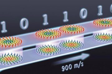
… a step towards the computing of the future. An international research team led by scientists from the CNRS1 has discovered that the magnetic nanobubbles2 known as skyrmions can be…


