Microscopy in the body
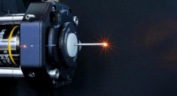
The endoscopy objective mounted on the coupling objective (Image: FAU/Sebastian Schürmann)
Laser used to illuminate molecules
It is often necessary to examine tissue samples under the microscope to diagnose diseases. This involves taking such samples using colonoscopy, for example, and applying contrast agents to distinguish different types of tissue effectively.
Biotechnologists, physicists and medical researchers at FAU have now developed a process that could greatly simplify examinations of the colon and other organs. They have miniaturised multi-photon microscopy to such an extent as to enable it to be used in endoscopes.
‘A multi-photon microscope emits focussed laser pulses at very high intensity for an extremely short period of time’, explains Prof. Dr. Dr. Oliver Friedrich from the Chair of Medical Biotechnology. ‘During this process, two or more photons interact simultaneously with certain molecules in the body which then makes the molecules illuminate.’
Multi-photon microscopy offers decisive advantages over conventional methods. Patients do not need to take synthetic contrast agents for the imaging of parts of connective tissue as the body’s own markers illuminate due to the excitation by photons.
In addition, the multi-photon laser penetrates deep into cells, for example into the walls of the colon, and provides high-resolution three-dimensional images of living tissue, whereas conventional colonoscopy is restricted to images of the surface of the colon. The procedure could supplement biopsies or even make them superfluous in some cases.
Multi-photon technology in a portable device
Multi-photon microscopes are already in use in medical applications, especially on the surface of the skin. For example, dermatologists use them to look for malignant melanoma. The challenge for using these microscopes in endoscopic examinations is the size of the technical components. Researchers at FAU have now successfully managed to house the entire microscope and femtosecond laser in a compact, portable device.
The objective lens is housed in a cannula that is 32 millimetres long and has a diameter of 1.4 millimetres. The focal point can be adjusted electronically to vary the optical penetration. A prism is located on the point of the needle allowing a sideways view into the colon, which means various rotational images of the tissue can be made from the same position.
In current experiments on small animals, the light emitted from the laser is transmitted via a rigid system. More research is needed to integrate the system into an endoscope. ‘Special photonic crystal fibres are required to guide the laser pulses’, says Friedrich.
‘Furthermore, in addition to the objective lens, the entire scanning mechanism must be miniaturised to allow it to be integrated into a flexible endoscope.’
Multi-photon organ atlas and pathologies
Multi-photon microendoscopy is not only useful for examining the colon. It could also be used in other areas of the body such as in the mouth and throat or in the bladder. The aim of the new method is to enable the doctor to detect whether organ cells and parts of the cell wall have changed on the micrometre scale. Complex dyeing processes and time-consuming biopsies can thus be limited. Prof. Friedrich’s team aim to provide doctors with an image database providing a multi-photon ‘atlas’ of organs and various diseases.
The project was funded by FAU’s Emerging Fields Initiative (EFI), which was set up in 2010. The aim of the initiative is to provide flexible funding to outstanding research projects as early as possible. Special consideration is given to interdisciplinary projects.
Further information:
Prof. Dr. Dr. Oliver Friedrich
Phone: +49 9131 8523174
oliver.friedrich@fau.de
Dr. Sebastian Schürmann
sebastian.schuermann@fau.de
DOI: 10.1002/advs.201801735: ‘Label-Free Multiphoton Endomicroscopy for Minimally Invasive In Vivo Imaging’
Media Contact
All latest news from the category: Life Sciences and Chemistry
Articles and reports from the Life Sciences and chemistry area deal with applied and basic research into modern biology, chemistry and human medicine.
Valuable information can be found on a range of life sciences fields including bacteriology, biochemistry, bionics, bioinformatics, biophysics, biotechnology, genetics, geobotany, human biology, marine biology, microbiology, molecular biology, cellular biology, zoology, bioinorganic chemistry, microchemistry and environmental chemistry.


Newest articles

High-energy-density aqueous battery based on halogen multi-electron transfer
Traditional non-aqueous lithium-ion batteries have a high energy density, but their safety is compromised due to the flammable organic electrolytes they utilize. Aqueous batteries use water as the solvent for…

First-ever combined heart pump and pig kidney transplant
…gives new hope to patient with terminal illness. Surgeons at NYU Langone Health performed the first-ever combined mechanical heart pump and gene-edited pig kidney transplant surgery in a 54-year-old woman…

Biophysics: Testing how well biomarkers work
LMU researchers have developed a method to determine how reliably target proteins can be labeled using super-resolution fluorescence microscopy. Modern microscopy techniques make it possible to examine the inner workings…

