Laser propelled cells
<br>
More than 40 years ago, the foundation for optical tweezers was laid when Arthur Ashkin demonstrated that near the focus of a laser beam, momentum transfer between light and dielectric particles creates gradient forces large enough to pull the particle into the center of highest intensity and scattering forces that push it in the propagation direction of the beam.
Optical trapping of microparticles and cells can be established either by balancing the axial forces of two weakly-focused counter-propagating beams or by using a single tightly focused laser beam. These optical tweezers have developed into an important tool in cell biological research. Optical tweezers can be used not only to fix cells during manipulation but also to investigate the interconnection of a cell’s elasticity to its physiology: healthy and diseased cells differ notably in their mechanical responses, prominent examples being blood disorders, asthma and cancer.
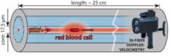
Researchers from Max Planck Institute for the Science of Light, Erlangen, Germany now report a new tool for biomechanical studies of individual cells: Single red blood cells were laser-propelled through stationary liquid in a microfluidic channel over distances of up to 24 cm. Shear forces on the cell surface result in its deformation. This causes changes in speed that can conveniently be monitored using a non-imaging laser Doppler-velocimetric technique. Numerical simulations allowed the scientists to derive the optical force acting on different cell shapes.
The unique method is based on a liquid-filled hollow-core photonic crystal fiber which provides low-loss light guidance in a well-defined single mode, resulting in highly uniform optical trapping and propulsive forces in the core which at the same time acts as a microfluidic channel. Cells are trapped laterally at the center of the core, several microns away from the glass interface, which eliminates adherence effects and external perturbations.
Dynamic changes in velocity at constant optical powers up to 350 mW indicated stress-induced changes in the shape of the cells, which was confirmed by bright-field microscopy. The deformations in the moving cells were not only due to heating. Even at moderate temperature, notable deformations could be detected, especially for osmotically swollen red blood cells.
Interestingly, the deformations occur over timescales of minutes which is rather slow compared to other cell rheological techniques. Re-arrangements of the cytoskeleton might be involved.
The scientists are currently aiming at studying suspended eukaryotic (cancer) cells. These cells are typically ellipsoidal in shape and more rigid than red blood cells, which prevents them from undergoing peculiar changes in shape.
Simulations of the optical forces would be possible, allowing for a complete theoretical analysis of the system. Beyond that, the method may find applications in on-chip cell transport. Cells might be held stationary against a mild counterflow carrying precise amounts of medical drugs.
Moreover, cell-cell interactions between suspended cells might be studied. (Text contributed by K. Maedefessel-Herrmann)
Unterkofler, S., et al; J. Biophotonics 6(9), 743-752 (2013); DOI http://dx.doi.org/10.1002/jbio.201200180
Journal of Biophotonics publishes cutting edge research on interactions between light and biological material. The journal is highly interdisciplinary, covering research in the fields of physics, chemistry, biology and medicine. The scope extends from basic research to clinical applications. Connecting scientists who try to understand basic biological processes using light as a diagnostic and therapeutic tool, the journal offers a platform where the physicist communicates with the biologist and where the clinical practitioner learns about the latest tools for diagnosis of diseases. JBP offers fast publication times: down to 20 days from acceptance to publication. Latest Journal Impact Factor (2012): 3.099 (ISI Journal Citation Reports 2012)
Regina Hagen
Journal Publishing Manager, Journal of Biophotonics
Managing Editor, Physical Sciences
Global Research
Wiley-VCH Verlag GmbH & Co. KGaA
Rotherstrasse 21
10245 Berlin
Germany
T +49 (0)30 47 031 321
F +49 (0)30 47 031 399
jbp@wiley.com
www.biophotonics-journal.org
www.wileyonlinelibrary.com
Media Contact
More Information:
http://www.wiley.comAll latest news from the category: Life Sciences and Chemistry
Articles and reports from the Life Sciences and chemistry area deal with applied and basic research into modern biology, chemistry and human medicine.
Valuable information can be found on a range of life sciences fields including bacteriology, biochemistry, bionics, bioinformatics, biophysics, biotechnology, genetics, geobotany, human biology, marine biology, microbiology, molecular biology, cellular biology, zoology, bioinorganic chemistry, microchemistry and environmental chemistry.


Newest articles

Superradiant atoms could push the boundaries of how precisely time can be measured
Superradiant atoms can help us measure time more precisely than ever. In a new study, researchers from the University of Copenhagen present a new method for measuring the time interval,…

Ion thermoelectric conversion devices for near room temperature
The electrode sheet of the thermoelectric device consists of ionic hydrogel, which is sandwiched between the electrodes to form, and the Prussian blue on the electrode undergoes a redox reaction…

Zap Energy achieves 37-million-degree temperatures in a compact device
New publication reports record electron temperatures for a small-scale, sheared-flow-stabilized Z-pinch fusion device. In the nine decades since humans first produced fusion reactions, only a few fusion technologies have demonstrated…

