Human gut microbiome physiology can now be studied in vitro using Organ Chip technology
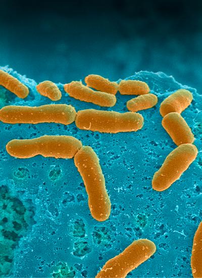
Bacteria of the human gut microbiome (yellow) are populating the intestinal epithelial channel of the anaerobic Intestine Chip that maintains gut-like low-oxygen concentrations. The bacteria are directly attaching to a dense mucus layer produced by the intestinal epithelium. Credit: Wyss Institute at Harvard University
The human microbiome, the huge collection of microbes that live inside and on our body, profoundly affects human health and disease. The human gut flora in particular, which harbor the densest number of microbes, not only break down nutrients and release molecules important for our survival but are also key players in the development of many diseases including infections, inflammatory bowel diseases, cancer, metabolic diseases, autoimmune diseases, and neuropsychiatric disorders.
Most of what we know about human-microbiome interactions is based on correlational studies between disease state and bacterial DNA contained in stool samples using genomic or metagenomic analysis.
This is because studying direct interactions between the microbiome and intestinal tissue outside the human body represents a formidable challenge, in large part because even commensal bacteria tend to overgrow and kill human cells within a day when grown on culture dishes. Many of the commensal microbes in the intestine are also anaerobic, and so they require very low oxygen conditions to grow which can injure human cells.
A research team at Harvard's Wyss Institute for Biologically Inspired Engineering led by the Institute's Founding Director Donald Ingber has developed a solution to this problem using 'organ-on-a-chip' (Organ Chip) microfluidic culture technology.
His team is now able to culture a stable complex human microbiome in direct contact with a vascularized human intestinal epithelium for at least 5 days in a human Intestine Chip in which an oxygen gradient is established that provides high levels to the endothelium and epithelium while maintaining hypoxic conditions in the intestinal lumen inhabited by the commensal bacteria.
Their “anaerobic Intestine Chip” stably maintained a microbial diversity similar to that in human feces over days and a protective physiological barrier that was formed by human intestinal tissue. The study is published in Nature Biomedical Engineering.
“The major paradigm shift in medicine over the past decade has been the recognition of the huge role that the microbiome plays in health and disease. This new anerobic Intestine Chip technology now provides a way to study clinically relevant human host-microbiome interactions at the cellular and molecular levels under highly controlled conditions in vitro,” said Ingber, M.D., Ph.D., is also the Judah Folkman Professor of Vascular Biology at Harvard Medical School (HMS) and the Vascular Biology Program at Boston Children's Hospital, and Professor of Bioengineering at SEAS.
“By providing direct access to the microbiome and differentiated intestinal tissue, this method can be used to discover specific microbes or their metabolites that cause disease or that might help prevent these conditions, and because we use cells isolated from patients, this approach could be used for personalized medicine as well.”
“Earlier tissue culture systems that aimed at recapitulating interactions between the human microbiome and intestinal epithelial cells in vitro were limited in their usefulness because they could not grow the two components in direct contact to one another, and did not mimic the gut's low oxygen concentrations crucial for the survival of anaerobic bacteria,” said first-author Sasan Jalili-Firoozinezhad, a Graduate Student shared between Ingber's team at the Wyss Institute and that of co-author Joaquim Cabral, Ph.D., Professor at the University of Lisbon, Portugal. Further complicating things: traveling along the small intestine towards the colon, oxygen levels continuously fall, which also changes the local microbiome composition.
For their anaerobic Intestine Chip, the team leveraged its proven Intestine Chip containing two parallel microchannels separated by a porous membrane. They grew human intestinal epithelial cells on the top of the membrane in the upper channel and vascular endothelial cells from intestinal microvessels on the opposite side of the membrane in the lower channel.
The intestinal cells used to line these Intestine Chips were either from a cell line or were obtained from human ileum biopsies and expanded through an intermediate organoid step in which they formed tiny, spherical intestinal tissue structures, which were broken up into fragments before being cultured in the chip.
To accommodate a complete microbiome, the team placed the Intestine Chips into a custom-engineered anaerobic chamber, that allowed them to drastically lower oxygen concentrations in the upper intestinal epithelial channel, while maintaining the lower endothelial channel at normal oxygen concentrations.
“We generated an oxygen gradient across the two channels that still allows the intestinal epithelium to be supported with oxygen diffusing through the porous membrane,” said co-first author Elizabeth Calamari, a Research Assistant on Ingber's team who engineered the device with Wyss Senior Staff Engineer and co-author Richard Novak, Ph.D. “In addition, we fitted the Intestine Chips with optical sensors that can report local oxygen concentrations in both channels in real-time without disturbing the oxygen gradient.”
Complex gut microbiome samples either obtained from healthy human stool and stably cultured in germ-free (gnotobiotic) mice or freshly isolated from infant stool, were then injected into the upper epithelial channel, where they came into direct contact with the mucus layer naturally secreted by the underlying intestinal epithelium. More importantly, the diversity of the commensal bacterial populations, when grown under these low-oxygen conditions, maintained the richness observed in human intestine.
“We showed through genome analysis we could culture over 200 distinct groups of bacteria for several days with abundances and ratios of obligate anaerobic bacteria similar to those observed in human stool,” said Jalili-Firoozinezhad. “Importantly, the complete microbiome further enhanced the barrier function of the intestinal epithelium with its cells providing a tight seal and producing a protective mucus layer, which is an important prerequisite for intestinal health.”
Being able to look at composition and changes of complete human microbiomes in direct contact with human intestinal tissue in vitro and over days opens up opportunities for personalized medicine and drug testing. “We can culture region-specific intestinal tissue and microbiomes from the same individual to find associations that cause sensitivity or tolerance to specific pathogenic, inflammatory, and systemic diseases,” said co-first author Francesca Gazzaniga, Ph.D., who is a Postdoctoral Fellow shared between Ingber's group and that of co-author Dennis Kasper, M.D., Professor of Immunology at HMS. “With the anaerobic Intestine Chip, we can also test the direct effects of drugs on the human microbiome before giving them to people.”
###
Other authors on the study are present and former members of Ingber's group and Wyss Institute staff, including Senior Informatics Scientist Diogo Camacho, Ph.D.; Clinical Fellow Cicely Fadel, M.D., Ph.D.; Postdoctoral Fellow Amir Bein, Ph.D.; Research Assistants Ben Swenor, Michael Cronce and Bret Nestor; Graduate Student Alessio Tovaglieri; former Staff Scientist Oren Levy, Ph.D., M.B.A; as well as Katherine Gregory, Ph.D., R.N., Executive Director for Women's and Newborn Health at Brigham and Women's Hospital; and David Breault, M.D., Ph.D., Associate Professor at HMS and Boston Children's Hospital. The study was funded by Harvard's Wyss Institute for Biologically Inspired Engineering, the Fundação para a Ciência e a Tecnologia (FCT) Portugal, the U.S. Federal Drug Administration, DARPA's Technologies for Host Resilience (THoR) initiative, and the Bill & Melinda Gates Foundation.
PRESS CONTACT
Wyss Institute for Biologically Inspired Engineering at Harvard University
Benjamin Boettner, benjamin.boettner@wyss.harvard.edu, +1 617-432-8232
MULTIMEDIA AVAILABLE
The Wyss Institute for Biologically Inspired Engineering at Harvard University uses Nature's design principles to develop bioinspired materials and devices that will transform medicine and create a more sustainable world. Wyss researchers are developing innovative new engineering solutions for healthcare, energy, architecture, robotics, and manufacturing that are translated into commercial products and therapies through collaborations with clinical investigators, corporate alliances, and formation of new startups. The Wyss Institute creates transformative technological breakthroughs by engaging in high risk research, and crosses disciplinary and institutional barriers, working as an alliance that includes Harvard's Schools of Medicine, Engineering, Arts & Sciences and Design, and in partnership with Beth Israel Deaconess Medical Center, Brigham and Women's Hospital, Boston Children's Hospital, Dana-Farber Cancer Institute, Massachusetts General Hospital, the University of Massachusetts Medical School, Spaulding Rehabilitation Hospital, Boston University, Tufts University, Charité – Universitätsmedizin Berlin, University of Zurich and Massachusetts Institute of Technology.
Media Contact
All latest news from the category: Life Sciences and Chemistry
Articles and reports from the Life Sciences and chemistry area deal with applied and basic research into modern biology, chemistry and human medicine.
Valuable information can be found on a range of life sciences fields including bacteriology, biochemistry, bionics, bioinformatics, biophysics, biotechnology, genetics, geobotany, human biology, marine biology, microbiology, molecular biology, cellular biology, zoology, bioinorganic chemistry, microchemistry and environmental chemistry.


Newest articles

Silicon Carbide Innovation Alliance to drive industrial-scale semiconductor work
Known for its ability to withstand extreme environments and high voltages, silicon carbide (SiC) is a semiconducting material made up of silicon and carbon atoms arranged into crystals that is…

New SPECT/CT technique shows impressive biomarker identification
…offers increased access for prostate cancer patients. A novel SPECT/CT acquisition method can accurately detect radiopharmaceutical biodistribution in a convenient manner for prostate cancer patients, opening the door for more…

How 3D printers can give robots a soft touch
Soft skin coverings and touch sensors have emerged as a promising feature for robots that are both safer and more intuitive for human interaction, but they are expensive and difficult…

