Deeper insight in the activity of cortical cells
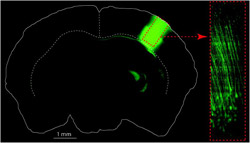
Left is an image of a cross-section through the whole mammalian brain that shows both brain hemispheres (solid white outline) as well as the overlaying cerebral cortex which is made up of many layers (I – VI). On the right hemisphere are brain cells, neurons, labeled with a genetically encoded fluorescent marker that reports back the cells activity by fast changes in brightness. This image has been taken from a brain slice post mortem where the lower limit of the cortex can be seen (dotted white line). Right, this image shows the same deep layer V brain cells (red box) labeled with the genetically encoded fluorescent marker but actually imaged non-invasively from a living animal using a modified multiphoton microscope, or RAMM approach. This allows scientists to study activity in neuronal populations deep in the cortex of an awake behaving animal and will lead to a deeper understanding of how cortical networks perform computations. © Wolfgang Mittmann, Jason Kerr / Max Planck Institute for Biological Cybernetics <br>
Visual and tactile objects in our surroundings are translated into a perception by complex interactions of neurons in the cortex. The principles underlying spatial and temporal organization of neuronal activity during decision-making and object perception are not all understood yet. Jason Kerr from Max Planck Institute for Biological Cybernetics in Tübingen, in collaboration with Winfried Denk from the Max Planck Institute for Medical Research in Heidelberg, now investigated how different sensations are represented by measuring activity in neuronal populations deep in the cortex. The scientists developed a method, with which they can study the neuronal activity in some of the deepest layers of the cortex in rodents, something that has not been possible up until now.
The cerebral cortex, or just cortex, is the outermost sheet of neural tissue of the mammalian brain. It plays a key role in memory, perceptual awareness and consciousness. It receives and processes the information from the senses, such as sight, touch or smell. The principles underlying these processes are not fully understood yet. Jason Kerr, research group leader of the Network Imaging Group at the Max Planck Institute for Biological Cybernetics in Tübingen and his colleagues from the same institute, Wolfgang Mittmann, Damian Wallace und Uwe Czubayko managed to image neuronal activity simultaneously from many neurons with single cell resolution, over twice as deep as previously achieved. In collaboration with Winfried Denk from the Department Biomedical Optics at the Max Planck Institute for Medical Research in Heidelberg and scientists from the Howard Hughes Medical Institute in Ashburn, Virginia they studied the neural cell activity in layer L5b in the adult rodent, which, as well as being one of the output layers of the cortex, it is also only one layer away from the cortex end.
Up until now. most imaging studies were restricted to the upper third of the cortex in the so-called layers L2 and L3. Deeper layers could only be studied using electrodes or by damaging the cortex using optical fibers or prisms. The Max Planck scientists now further developed a method, with which they can see exactly which cell is active and importantly, what cells are not active during a stimulus up to one millimeter from the cortical surface. This has enabled the scientists to measure the spatiotemporal organization of activity in these deep layers.
“We express a genetically encoded fluorescent activity reporter in the neurons of interest and with this we can measure the activity of many neurons at the same time”, explains Jason Kerr. Changes in brightness of the fluorescent marker are relative to the activity of the neuron. Using the new multi photon imaging technique the activity of many neuronal populations in the deeper cortical layers can be recorded simultaneously in vivo. Jason Kerr and his team combined regenerative amplification multiphoton microscopy (RAMM) with generally encoded calcium indicators to extend multi photon imaging of neuronal population activity to the deeper layers of the cortex. Using this approach, they found, that it could be used to record and quantify spontaneous and activity evoked in the animal by sensory stimulation such as whisker touches or natural movies in neuronal populations of the layer L5a and L5b.
The goal of their research is to record activity from populations of neurons located in all cortical layers, from the layer 6 to layer 1. In combination with genetically encoded activity indicators, the team plans to investigate the spatial temporal organization of neuronal activity from all cortical layers in animals trained to discriminate between objects. Further, they want to address the question of whether the deeper layers also show spatiotemporal re-organization similar to that shown for the upper cortical layers during learning. With these technical advances the scientists aim to gain insights into cortical circuits involved in decision making in the awake, behaving cortex, and how these circuits are functionally modified during learning.
Contact
Dr. Jason Kerr
Max Planck Institute for Biological Cybernetics, Tübingen
Phone: +49 70 7160-11721
Email: Jason@tuebingen.mpg.de
Stephanie Bertenbreiter
Press and Public Relations
Max Planck Institute for Biological Cybernetics, Tübingen
Phone: +49 7071 601-472
Email: presse@tuebingen.mpg.de
Original publication
Wolfgang Mittmann, Damian J Wallace, Uwe Czubayko, Jan T Herb, Andreas T Schaefer, Loren L Looger, Winfried Denk & Jason N D Kerr.
(2011) Two-photon calcium imaging of evoked activity from L5 somatosensory neurons in vivo.
Nature Neuroscience doi:10.1038/nn.2879
Media Contact
More Information:
http://www.mpg.de/4369894/cortical_cell_activityAll latest news from the category: Life Sciences and Chemistry
Articles and reports from the Life Sciences and chemistry area deal with applied and basic research into modern biology, chemistry and human medicine.
Valuable information can be found on a range of life sciences fields including bacteriology, biochemistry, bionics, bioinformatics, biophysics, biotechnology, genetics, geobotany, human biology, marine biology, microbiology, molecular biology, cellular biology, zoology, bioinorganic chemistry, microchemistry and environmental chemistry.


Newest articles

Silicon Carbide Innovation Alliance to drive industrial-scale semiconductor work
Known for its ability to withstand extreme environments and high voltages, silicon carbide (SiC) is a semiconducting material made up of silicon and carbon atoms arranged into crystals that is…

New SPECT/CT technique shows impressive biomarker identification
…offers increased access for prostate cancer patients. A novel SPECT/CT acquisition method can accurately detect radiopharmaceutical biodistribution in a convenient manner for prostate cancer patients, opening the door for more…

How 3D printers can give robots a soft touch
Soft skin coverings and touch sensors have emerged as a promising feature for robots that are both safer and more intuitive for human interaction, but they are expensive and difficult…

