Dancing with the cells
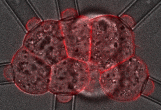
The scientists measured the force required to change the shape of the cells by gently deforming each cell with a pipette. IMAGE: J.L. Maitre/EMBL
The same kind of contraction that fires our muscles also controls a key stage of mammalian embryo development, according to a new study published in Nature Cell Biology. The research, conducted at EMBL Heidelberg, measured and mapped how cells in very early stage embryos bond tightly together. The scientists also discovered a cellular behaviour that hadn’t been observed before: cells in the embryo ‘dance’, each one making the same rhythmic movement.
The focus of the study was a stage of development known as compaction, which takes place when the embryo has eight cells. Compaction changes the embryo from a loosely attached group of cells to a closely bonded single entity. During compaction – which takes around 10 hours – the cells change shape to create the overall form of the embryo, increasing the area of contact between them.
Using a new method, the researchers were able to measure the forces required to change the shape of the cells as compaction progressed.
Being able to chart the tension within the embryo without destroying it meant they were able to investigate which cellular process was the main driver behind the compaction process.
The first contender was a process known as adhesion. This is controlled by E-cadherin, an adhesion molecule on the cell surface that sticks cells together, ’zipping up’ the two surfaces as the molecules attach to each other. Earlier research had shown that when adhesion was blocked, compaction did not take place.
The second contender was cell contraction, a process controlled by myosin, a type of motor protein that also causes contractions in muscle fibres. Myosin ‘walks’ on tracks formed by another protein, actin. Every cell has a layer of actin underneath its membrane, and myosin contracts this cortical layer, controlling the tension of the cell surface. The EMBL Heidelberg team had also established that compaction did not take place if a cell’s ability to contract was blocked.
Postdoctoral researcher and first author, Jean-Léon Maître, explains: “By measuring the tensions of the cells when each cellular process was blocked, we were able to prove it was a contraction that pulls the cells together to compact the embryo, rather than adhesion acting as a glue to ‘stick’ them together. Adhesion is obviously important, but it appears to work as an anchor, rather than an engine of the compaction process.”
This discovery was made possible by combining biological expertise from the group led by Takashi Hiiragi with that of the physicists working in the Nédéléc group.
Hervé Turlier explains: “The research showed that it was the increased tension at the outer surface of the embryo that drives the compaction, rather than the relaxation of the cell to cell contacts. In fact, the changing ratio between these two tensions now provides us with a simple way to portray the compaction process despite the complex biological mechanisms taking place.”
At the same time as compaction but on a shorter timescale, the team observed that the cells start to ‘dance’. The ‘dance’ is caused by a wave of contraction that bends the surface of the cell, traveling round it every 80 seconds.
“We’ve no idea at the moment if this ‘dance’ is important,” says Hiiragi. “All we know is that it happens at the same time as compaction and is controlled by the same process.”
Embryonic cells in other animals are known to pulse every 80 seconds, but this particular form of the movement hasn’t been observed before. Further research will hopefully unveil the details underlying this peculiar phenomenon.
Published online in Nature Cell Biology on 15 June 2015. DOI: 10.1038/ncb3185.
For images, video and more information please visit:
Media Contact
All latest news from the category: Life Sciences and Chemistry
Articles and reports from the Life Sciences and chemistry area deal with applied and basic research into modern biology, chemistry and human medicine.
Valuable information can be found on a range of life sciences fields including bacteriology, biochemistry, bionics, bioinformatics, biophysics, biotechnology, genetics, geobotany, human biology, marine biology, microbiology, molecular biology, cellular biology, zoology, bioinorganic chemistry, microchemistry and environmental chemistry.


Newest articles

Security vulnerability in browser interface
… allows computer access via graphics card. Researchers at Graz University of Technology were successful with three different side-channel attacks on graphics cards via the WebGPU browser interface. The attacks…
A closer look at mechanochemistry
Ferdi Schüth and his team at the Max Planck Institut für Kohlenforschung in Mülheim/Germany have been studying the phenomena of mechanochemistry for several years. But what actually happens at the…

Severe Vulnerabilities Discovered in Software to Protect Internet Routing
A research team from the National Research Center for Applied Cybersecurity ATHENE led by Prof. Dr. Haya Schulmann has uncovered 18 vulnerabilities in crucial software components of Resource Public Key…

