Automated analysis of whole brain vasculature
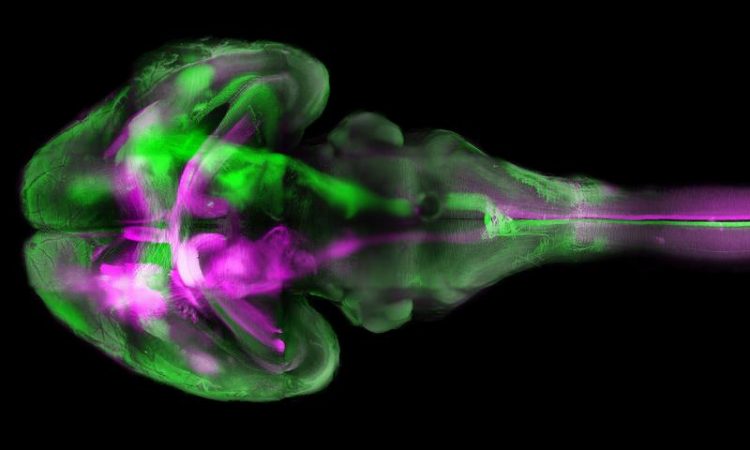
TThe brain of a mouse - taken with fluorescence microscopy using "tissue clearing" - a technique that for the first time made the large and small brain vessels visible simultaneously. Ertürk Lab / Institute for Stroke and Dementia Research
Changes in the blood vessels are a hallmark of numerous brain disorders – from traumatic brain injury to stroke. Even diseases such as Alzheimer's show changes in the fine capillaries. In short, analysing the blood vessels is key to understanding both normal and pathological brain function.
“Now we have come much closer to achieving that goal”, explains Ali Ertürk, Director of the Institute for Tissue Engineering and Regenerative Medicine at Munich's Helmholtz Centre and Principal Investigator at the Institute for Stroke and Dementia Research at the LMU University Hospital Munich.
Making organs transparent
As a first step, Ertürk's team succeeded in visualising the vascular system of mouse brains with high-resolution fluorescent microscopy without having to cut up the specimens into small sections.
In order to do this, they refined the technique of tissue cleaning, in which biological tissues are treated with special dyes to render them transparent for fluorescent microscopy. “Previously, this technique could only be used to scan either the large vessels of the brain or the small ones”, says Mihail Ivilinov Todorov, a doctoral student studying under Ertürk.
Therefore the Munich-based scientists took the new approach of combining two dyes. “That gave us some great images of the brain vasculature including the capillaries”, adds the biologist.
Vascular network captured using artificial intelligence
Applying artificial intelligence, researchers from the team led by Björn Menze, Professor for Machine Learning in Biomedical Imaging at the Technical University of Munich, used these images to reconstruct the entire vascular network of the brain right down to its finest details.
Such a reconstruction yields more than just images – it also allows a quantitative analysis of the vascular structures. “For example, we can statistically record the diameters of the various blood vessels or their bifurcations for different areas of the brain”, says Johannes Paetzold, doctoral student in Menze’s group.
“Over the past few years, we have developed a deep learning algorithm that specialises in detecting blood vessels in medical images”, Menze explains. “This was the first time we applied it to a whole brain.” The algorithm was able to reliably distinguish between blood vessels and other tissue even though some areas in the original fluorescence images were not well-illuminated and some details were distorted due to light reflections or other errors.
Understanding and diagnosing brain disorders
Mihail Ivilinov Todorov plans to use the statistical data in order to investigate vascular changes caused by stroke, while Björn Menze is looking to study the global structures of the vascular system in order to understand the role of anatomical differences in brain disorders, for example.
Benefits for the patient
The method could also be used in everyday clinical practice: “With our system, we are likely to be able to analyse the small tissue specimens from human tumours with greater accuracy”, Ertürk asserts. Cancerous tissue is permeated by blood vessels, and analysing their structure helps in staging a tumour.
“This may have an optimising effect on treatment”, Ertürk adds. The biologist also plans to use the new method to realise his vision for the future: the production of human organs on a 3D printer. For that to happen, a knowledge of the organ's precise vascular structure – among many other things – will be vital.
Dr. Ali Ertürk
Helmholtz Centre Munich /
Principal Investigator at the Institute for Stroke and
Dementia Research at the LMU University Hospital Munich
Email: ali.ertuerk@med.uni-muenchen.de
Web: https://www.isd-research.de/erturk-lab
Prof. Dr. Björn Menze
Technical University of Munich (TUM)
Professor for Machine Learning in Biomedical Imaging
Munich School of BioEngineering and Central Institute for
Translational Cancer Research (TranslaTUM)
Phone: +49 89 289 10930
Email: bjoern.menze@tum.de
Media Contact
All latest news from the category: Life Sciences and Chemistry
Articles and reports from the Life Sciences and chemistry area deal with applied and basic research into modern biology, chemistry and human medicine.
Valuable information can be found on a range of life sciences fields including bacteriology, biochemistry, bionics, bioinformatics, biophysics, biotechnology, genetics, geobotany, human biology, marine biology, microbiology, molecular biology, cellular biology, zoology, bioinorganic chemistry, microchemistry and environmental chemistry.


Newest articles

Bringing bio-inspired robots to life
Nebraska researcher Eric Markvicka gets NSF CAREER Award to pursue manufacture of novel materials for soft robotics and stretchable electronics. Engineers are increasingly eager to develop robots that mimic the…

Bella moths use poison to attract mates
Scientists are closer to finding out how. Pyrrolizidine alkaloids are as bitter and toxic as they are hard to pronounce. They’re produced by several different types of plants and are…

AI tool creates ‘synthetic’ images of cells
…for enhanced microscopy analysis. Observing individual cells through microscopes can reveal a range of important cell biological phenomena that frequently play a role in human diseases, but the process of…

