New ultra-miniaturized scope less invasive, produces higher quality images
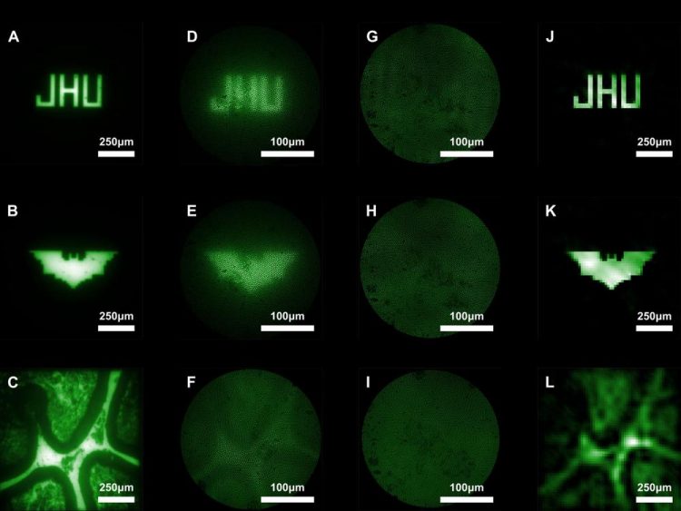
A to C shows beads on a slide imaged by a bulk microscope. D to F are the beads when viewed from a conventional lens-based microendoscope. G to I are the raw images from the research team's new ultra-miniaturized lens-free microendoscope. The researchers say these images are terrible but actually provide a great deal of information about light coming through that can be used in computational reconstruction to piece together a clearer final image. J to L are images G to I after computational reconstruction. Credit: Mark Foster
“Usually, you have sacrifice either size or image quality. We've been able to achieve both with our microendoscope,” says Mark Foster, an associate professor of electrical and computer engineering at The Johns Hopkins University and the study's corresponding author.
Intended for examining neurons firing off in the brains of animals such as mice and rats, an ideal microendoscope should be small to minimize brain tissue damage yet powerful enough to produce a clear image.
Currently, standard microendoscopes are about half a millimeter to a few millimeters in diameter, and require larger, more invasive lenses for better imaging. While lensless microendoscopes exist, the optical fiber within that scans an area pixel by pixel frequently bends and loses imaging ability when moved.
In their new study, Foster and colleagues created a lens-free ultra-miniaturized microendoscope that, compared to a conventional lens-based microendoscope, increases the amount researchers can see and improves image quality.
The researchers achieved this by using a coded aperture, or a flat grid that randomly blocks light creating a projection in a known pattern akin to randomly poking a piece of aluminum foil and letting light through all of the small holes.
This creates a messy image, but one that provides a bounty of information about where the light originates, and that information can be computationally reconstructed into a clearer image. In their experiments, Foster's team looked at beads in different patterns on a slide.
“For thousands of years, the goal has been to make an image as clear as possible. Now, thanks to computational reconstruction, we can purposefully capture something that looks awful and counterintuitively end up with a clearer final image,” says Foster.
Additionally, Foster and team's microendoscope doesn't require movement to focus on objects at different depths; they use computational refocusing to determine where the light originated from in 3 dimensions. This allows the endoscope to be much smaller than a traditional one that requires moving the endoscope around to focus.
Looking forward, the research team will test their microendoscope with fluorescent labeling procedures in which active brain neurons would be tagged and illuminated, to determine how accurately the endoscope can image neural activity.
###
Other authors on the study include Jaewook Shin, Dung N. Tran, Jasper R. Stroud, Sang Chin and Trac D. Tran, all of The Johns Hopkins University.
Media Contact
All latest news from the category: Information Technology
Here you can find a summary of innovations in the fields of information and data processing and up-to-date developments on IT equipment and hardware.
This area covers topics such as IT services, IT architectures, IT management and telecommunications.


Newest articles

Bringing bio-inspired robots to life
Nebraska researcher Eric Markvicka gets NSF CAREER Award to pursue manufacture of novel materials for soft robotics and stretchable electronics. Engineers are increasingly eager to develop robots that mimic the…

Bella moths use poison to attract mates
Scientists are closer to finding out how. Pyrrolizidine alkaloids are as bitter and toxic as they are hard to pronounce. They’re produced by several different types of plants and are…

AI tool creates ‘synthetic’ images of cells
…for enhanced microscopy analysis. Observing individual cells through microscopes can reveal a range of important cell biological phenomena that frequently play a role in human diseases, but the process of…

