Multiple sclerosis
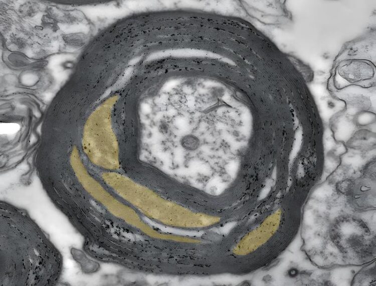
The figure shows an electron micrograph of a myelinated nerve fiber in the optic nerve of an MS patient. It can be seen that the myelin sheath contains loosened areas (colored yellow). The black bar corresponds to 0.5 micrometers.
Credit: Wiebke Möbius / Max Planck Institute for Multidisciplinary Sciences
Ultrastructural changes in brain tissue promote inflammatory processes.
Multiple sclerosis (MS) is the most common inflammatory disease of the central nervous system. It is characterized by inflammatory foci and damage in the brain’s so-called white matter, which consists of long nerve fibers and myelin. A German-Dutch research team has now shown that ultrastructural changes in healthy areas in the white matter of MS patients make the tissue more susceptible to inflammation and the formation of lesions. This could promote the progression of the disease.
It is 7 am, the alarm clock rings – you open your eyes, swing your legs out of bed, give a talk at work, play a tennis match in the evening. Billions of nerve cells that make up the gray matter in our brain allow us to perform these different tasks. They are interconnected millions of times by nerve fibers running deeper in the brain, called axons. Many of these axons are wrapped by a cellular “insulating tape”. The insulating cover is made of myelin, a lipid-rich substance that coats axons in up to 150 layers. Together, axons and myelin form what is known as white matter. At regular intervals, the myelin sheaths have a small gap called a node of Ranvier. When a signal is transmitted from one cell to the other by means of an electrical nerve impulse, it literally jumps from one node to the next. This accelerates communication on myelinated axons by 100 times.
The white matter, however, is more than a well-sorted and optimally insulated “cable collection”. It helps with various different processes in the brain, such as learning, memory, or social skills. If this tissue is damaged, diseases such as MS can develop. In MS, which affects up to 280,000 people in Germany alone, the myelin layer around the axons is damaged or destroyed. These lesions, combined with inflammatory reactions, can be detected by imaging techniques.
However, scientists still know little about how changes in axons and myelin at the subcellular level are related to inflammatory processes. Researchers led by Wiebke Möbius at the Max Planck Institute (MPI) for Multidisciplinary Sciences in Göttingen and Inge Huitinga at the Netherlands Institute for Neurosciences in Amsterdam (the Netherlands) have now discovered that the fine structure – called ultrastructure – of the seemingly normal white matter in MS patients is already altered, before the first foci of inflammation appear.
Quality of myelin samples decisive
“The areas that may be critical for MS disease can only be studied at the ultrastructural level using electron microscopy,” Möbius says. However, she adds, the tissue structures are often damaged in conventional preparation methods due to chemical fixation and embedding. “This is especially true for the sensitive myelin. The big challenge for us, therefore, was to preserve the structures in the tissue sample better than before. To do this, we optimized the fixation method of the samples in particular,” explains the head of the Facility for Electron Microscopy at the MPI’s City Campus.
For their experiments, the scientists used tissue samples from the Netherlands Brain Bank. These came from MS patients who had agreed during their lifetime to donate their brains post-mortem for research and medical records.
As the team showed, in the normal appearing white matter of MS patients, the myelin sheaths are conspicuously altered and the myelin is less compact. The nodes of Ranvier are also disorganized. In addition to these structural changes, the researchers found cellular markers for inflammation in the apparently normal tissue: T lymphocytes and activated immune cells of the brain called microglial cells. Last but not least, the density of mitochondria – the power generators of the cell – was strikingly increased in the processes of nerve cells, suggesting that communication between nerve cells requires more energy than in healthy people. Aletta van den Bosch from the Dutch group explains: “Mitochondria not only produce vital energy, but also many by-products such as oxygen radicals. We suspect that these may increase myelin damage.”
Möbius adds: “We can see clearly that in MS, ultrastructural changes in the white matter are related to chronic inflammation in the brain. Both pathological abnormalities could contribute to the progression of the disease. The fact that the ultrastructure of myelin can now be preserved in such quality for studies under the electron microscope will hopefully provide further new insights.”
Contact for scientific information:
Dr. Wiebke Möbius
Head of the Facility for Electron Microscopy
Max Planck Institute for Multidisciplinary Sciences (City Campus), Göttingen, Germany
phone: +49 551 201-31786
e-mail: moebius@mpinat.mpg.de
Original publication:
van den Bosch, A. M. R.; Hümmert, S.; Steyer, A.; Ruhwedel, T.; Hamann, J.; Smolders, J.; Nave, K.-A.; Stadelmann, C.; Kole, M. H. P.; Möbius, W.; Huitinga, I.: Ultrastructural axon-myelin unit alterations in multiple sclerosis correlate with inflammation. Ann Neurol 93, 856-870 (2022).
https://doi.org/10.1002/ana.26585
More information:
https://www.mpinat.mpg.de/4456755/pr_2306 – Original press release
https://www.mpinat.mpg.de/em-moebius – Website of the Facility for Electron Microscopy (City Campus), Max Planck Institute for Multidisciplinary Sciences, Göttingen, Germany
Media Contact
All latest news from the category: Life Sciences and Chemistry
Articles and reports from the Life Sciences and chemistry area deal with applied and basic research into modern biology, chemistry and human medicine.
Valuable information can be found on a range of life sciences fields including bacteriology, biochemistry, bionics, bioinformatics, biophysics, biotechnology, genetics, geobotany, human biology, marine biology, microbiology, molecular biology, cellular biology, zoology, bioinorganic chemistry, microchemistry and environmental chemistry.


Newest articles

Zap Energy achieves 37-million-degree temperatures in a compact device
New publication reports record electron temperatures for a small-scale, sheared-flow-stabilized Z-pinch fusion device. In the nine decades since humans first produced fusion reactions, only a few fusion technologies have demonstrated…
Innovative microscopy demystifies metabolism of Alzheimer’s
Researchers at UC San Diego have deployed state-of-the art imaging techniques to discover the metabolism driving Alzheimer’s disease; results suggest new treatment strategies. Alzheimer’s disease causes significant problems with memory,…
A cause of immunodeficiency identified
After stroke and heart attack: Every year, between 250,000 and 300,000 people in Germany suffer from a stroke or heart attack. These patients suffer immune disturbances and are very frequently…

