Embryonic development: How do limbs develop from cells?
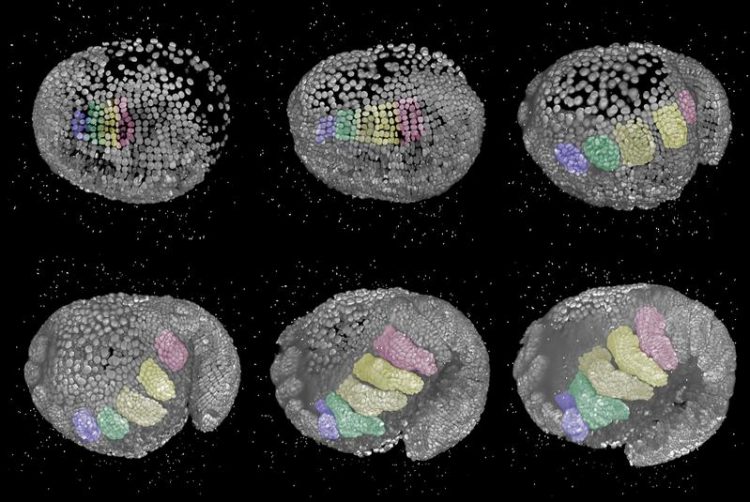
The developmental sequence of Parhyale hawaiensis captured using alight screen fluorescence microscope. This concerns 3D reconstructions of an embryo from a lateral view and the head on the left side. The areas highlighted in colourshow how legs differ on the basis of just a small number of cells. Copyright owners: Anastasios Pavlopoulos and Carsten Wolff
During the development of a multicell organism, a single cell produces a range of different cell types which in turn form various tissues and organs.
For over a century, biologists have been attempting to understand the biogenesis (also known as cell fate) of the different structures in developing organisms. For instance, which cells produce eyes or limbs? How does the behaviour of these developing cells change in order to realise the form and function of the respective organ?
Until now, it has been exceedingly difficult to answer these kinds of questions since directly observing developing embryos at the cellular level poses a considerable challenge.
Following a new approach, a multinational and interdisciplinary team of scientists from the Humboldt University of Berlin, the Max-Planck Institute for Molecular Cell Biology and Genetics (Dresden), the HHMI Janelia Research Campus (USA), the Max-Delbrück Centre for Molecular Medicine (Berlin) and the Pasteur Institute (France) is investigating the dynamics and behaviour of cells during limb development.
In this connection, the research team is taking a closer look at the development of the marine crustacean Parhyale hawaiensis. This creature has developed an array of specialised limbs along the length of its body – resembling a living Swiss army knife.
Using state-of-the-art light sheet fluorescence microscopy, the researchers were able to record the entire embryonic development of embryos with fluorescent cell nuclei. This procedure involved the embryos being observed from different perspectives in a high spatial and temporal resolution.
Cell monitoring can provide insights into deformities
Having collecting these complex and multidimensional microscopic images, the researchers faced the challenge of extracting the relevant biological information from these huge data sets. To this end, they developed a special software program called Massive Multi-View Tracker (MaMuT), which is freely available as a FIJI plug-in. This combination of microscopy and software allowed the scientists to examine all the cells of a leg during development over the course of several days.
They were thus able to discover morphogenetic behavioural patterns of the cells which contribute to the cellular organisation and growth of a leg. In the future, this new method of examining the development of multicell organisms will help to identify the cellular and molecular mechanisms that determine the fate of cells during the development of an organism. It will also undoubtedly prove useful in explaining the causes of deformities during development.
Article
https://elifesciences.org/articles/34410
Foto description
The developmental sequence of Parhyale hawaiensis captured
using alight screen fluorescence microscope. This concerns 3D
reconstructions of an embryo from a lateral view and the head
on the left side. The areas highlighted in colourshow how legs differ
on the basis of just a small number of cells.
Contact
Dr Carsten Wolff
Institute for Biology
Tel.: 030 2093-6284
carsten.wolff@rz.hu-berlin.de
Media Contact
All latest news from the category: Life Sciences and Chemistry
Articles and reports from the Life Sciences and chemistry area deal with applied and basic research into modern biology, chemistry and human medicine.
Valuable information can be found on a range of life sciences fields including bacteriology, biochemistry, bionics, bioinformatics, biophysics, biotechnology, genetics, geobotany, human biology, marine biology, microbiology, molecular biology, cellular biology, zoology, bioinorganic chemistry, microchemistry and environmental chemistry.


Newest articles

Superradiant atoms could push the boundaries of how precisely time can be measured
Superradiant atoms can help us measure time more precisely than ever. In a new study, researchers from the University of Copenhagen present a new method for measuring the time interval,…

Ion thermoelectric conversion devices for near room temperature
The electrode sheet of the thermoelectric device consists of ionic hydrogel, which is sandwiched between the electrodes to form, and the Prussian blue on the electrode undergoes a redox reaction…

Zap Energy achieves 37-million-degree temperatures in a compact device
New publication reports record electron temperatures for a small-scale, sheared-flow-stabilized Z-pinch fusion device. In the nine decades since humans first produced fusion reactions, only a few fusion technologies have demonstrated…

